Showing posts with label cancer. Show all posts

Tumor Lysis Syndrome (Truong, 11/19/2025)

 Thanks so much to our Pharmacy Resident, Lam Truong, who gave an excellent Grand Rounds this week on Tumor Lysis Syndrome.

Recording will be posted HERE when available. 

In the meantime, enjoy the cliff notes (thanks Cherie Green!)

Tumor Lysis Syndrome

  • Oncological emergency from rapid breakdown of malignant cells leading to massive release of intracellular contents into bloodstream
  • Overall in hospital mortality is 21%
  • Most common in NHL(30%) > solid tumors (20%) > AML (19%)> ALL (13%)
  • TLS can either present at initial dx with very aggressive tumors (usually lymphomas/leukemias but occasional very aggressive solid tumors), OR onset can be 3-7 days after chemotherapy - so patients with upcoming chemotherapy get labs 3 days prior to chemotherapy both to make sure they are not already in TLS and to compare post-chemo labs


Diagnosis: Prompt recognition and tx are essential….

4 Lab abnormalities for dx: 

Hyperkalemia (tumor cell lysis), Hyperphosphatemia (cell lysis), Hyperuricemia (from breakdown of DNA/RNA-crystal nephropathy), and Hypocalcemia (binds the excess phosphate)

Result:AKI (uric acid crystals, direct cytotoxicity), cardiac arrhythmias (hyperK), and seizures (metabolic disturbances), neuromuscular dysfunction (rigidity)

Symptoms: n/v/d, weakness, muscle cramps, paresthesias, seizures, arrhythmia, hypotension, HF, syncope, oliguria, hematuria, edema, joint pain - so keep your differential broad if patients present with these sxs!


Causes: chemo causing cytotoxic effects, molecular targeted therapies and immunotherapies, can occur spontaneously in setting of large tumor burden even in absence of treatment initiation


Monitor Uric acid, K Phos, Ca, Cr, LDH (high LDH represents rapid cell turnover)


Prevention of Tumor Lysis Syndrome:

Hydration protocol, closely monitor UO, dc nephrotoxins, stop all K agents 

Hypouricemic Agents for prevention and tx

Allopurinol xanthine oxidase inhibitor used in intermediate risk patients, prophylaxis in TLS but has slower onset than…

Rasburicase recombinant urate oxidase inhibitor for high risk patients and tx of active hyperuricemia works rapidly (risks: hemolytic anemia in G6PD deficiency). Loading dose 3 mg IV x 1 


Management: Use Tumor Lysis Order Set 

  • Look at criteria and lab monitoring - some need additional doses of rasburicase and q6 hr labs
  • HyperK: use protocol
  • Hyperphos: treat first with binders and concurrently treat hypocalcemia
  • HypoCa: look on the line above, don't treat first. Treat Phos first!
  • HD for usual indications (refractory hyperK, fluid overload, etc)


Anal Cancer Detection (Mejia Powell, 9/17/25)

 A recording of this presentation is available HERE.

Dr. Rob Mejia Powell gave a really awesome talk this week about Anal Cancer Screening. He reminded us that, while anal squamous cell carcinoma-- almost entirely HPV-related -- is rare in the overall population (1-2 cases/100K person years). While most people are not high risk and should NOT be screened, there are higher risk populations who should definitely be screened with anal pap smears and referred to high resolution anoscopy (HRA), which is available through SRCH for internal and external referrals. Take a look at his presentation for some really great info. If you just want the brief notes, you'll miss out on the butt jokes. 

Here's a reminder of what patients with anal cancer "look like", survivors of anal cancer:   https://youtu.be/QYR3GWWAmjE?si=kolJuD1TrdgWXMBU

"Say the word 'anal anal anal' a million times until your friends get used to it."

Take a look at this table to see the relative incidence in certain risk categories:

Known risk factors include age, HPV infection (especially vulvar neoplasia in women), receptive anal intercourse, and immunosuppression (e.g. HIV, chronic steroid/immunosuppressants). Also note that the highest growing group in women.

Pathogenesis of anal cancer is similar to cervical cancer:  Exposure to HPV > persistent infection > precancerous high-grade squamous intraepithelial lesions (HSIL, anal intraepithelial neoplasia (AIN grades 2 or 3)) > invasion to ASCC


Who should be screened?

  • People with HIV
  • MSM (especially with HIV)
  • Women with history of cervical/vulvar/vaginal neoplasia
  • Transgender women
  • Solid organ transplant recipients, IBD (esp perianal Crohn's) and those on chronic immunosuppressants
Does screening work?
The ANCHOR Trial, published in NEJM 2022 was a multi-site RCT that showed that, YES, screening at risk populations does work to prevent anal cancer. Primary outcome: anal cancer. Test subjects: Men with HIV age >35. Study found 9 anal cancers in the treatment group (those screened>> HRA>> treatment) vs. 21 cancers in the observation group. This equates to a 60% decrease in anal cancer.

How to approach the topic of anal cancer screening with patients:
1) Explain what an anal pap smear is: what are we looking for? what are we trying to prevent? 
2) Get an HPI: anal itching? bleeding? pain? difficulty stooling? presence of anal lesions?
3) Anal Pap: get cytology and HPV prior to digital exam>> use Dacron swab (wet with water, place 2-3 inches across anal verge, aggressive angling)
4) Digital anal rectal exam (DARE)
5) Refer abnormal anal paps to high res anoscopy (HRA) (see chart)

Summary chart: 


Colorectal Cancer Screening (Toub, 2/14/2024)

 A recording of this presentation is available HERE

***

Many thanks to Dr. Danny Toub, who gave an excellent Grand Rounds presentation this week titled Colorectal Cancer Screening: What PCPs need to Know. A recording of the presentation is available above. 

My notes:

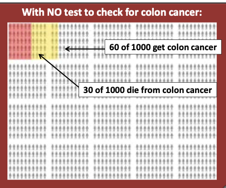
Colorectal cancer (CRC) is the #2 cause of US cancer deaths overall in men and women, the #1 cause of cancer death in men under 50. In 2024, we are predicted to have more CRC deaths than COVID deaths. In theory, CRC is an ideal disease to screen for because the slow development from polyp to cancer -- on average, 10 years -- means that early detection can actually lead to lives saved; if caught early, death can be prevented. 

Through many efforts, we have increased our CRC screening over the last two decades, and in doing so, we have  decreased CRC mortality. Unfortunately, it's not by a lot: a 2023 study published in JAMA, found that CRC screening only extends a person's life by an average of 110 days (see forest plot below). 

Despite what seems like a perfect set-up for screening success (a slow growing cancer with multiple tools for early detection), the reality is that CRC screening doesn't have great evidence for all-cause mortality benefit either. Sigh. Plus, the number needed to screen (NNS) to prevent one cancer death is not small -- 450 with q5 year flex sig and 900 with annual FOBT. Double sigh. 

It is important to note, however, that current quality measures do incentivize us to screen for CRC-- there are payments attached to our doing so, through both state and federal programs. So, to be clear, we screen to 1) decrease CRC mortality and 2) because our systems are set up to do so. 

How do we screen? 

Many of us are well aware that there are several CRC screening modalities, which are essentially considered equivalent in terms of CRC detection rates:

  1. q10 year colonoscopy (most invasive, most cumbersome, but benefit of being able to remove polyps during the procedure)
  2. Annual FIT test (has replaced the older FOBT test)
  3. stool DNA tests (frequency variable)
  4. q5 year flexible sigmoidoscopy (screens less of the colon but does not require anesthesia and can be done by primary care clinicians, if trained)
  5. Emerging serum tests (not yet FDA approved)

Current USPSTF Guidelines (2021) recommend shared decision-making to decide which modality to use; there are a number of studies showing many different screening tools decrease CRC mortality (with colonoscopy slightly better than the other modalities in detecting cases and averting death). More invasive screening tools have more serious potential harms, but all positive screening tests lead to colonoscopy, as the gold standard for diagnosis of CRC. Of note, there is a lack of screening colonoscopy access in our county, and most of the community health centers (and even Kaiser) default to non-invasive screening via FIT testing. As an aside, SRCH is moving to a pilot program screening with a more expensive DNA FIT testing q3 year (rather than q year) starting next month. 

Who to screen?

Current USPSTF Guidelines (2021) make a Grade A recommendation for all adults 50-75, with a Grade B recommendation for adults 45-49. They make a Grade C recommendation for people 76-85 with shared decision-making based on 10 year life expectancy (i.e. healthy 76+ year adults should  be selectively screened).

Whereas there is a statistical benefit to screening people 45-49, the AAFP actually disagrees with the USPSTF and recommends  screening starting age 50 with "insufficient evidence (I)" to recommend screening before then. The 2023 ACP guidelines are slightly less in terms of screening frequency, recommending a q2 year FIT test (vs 1 year per USPSTF).

What works?

Dr. Toub spent some time talking about how we get patients to get screened -- possible interventions include one on one conversations, client reminders, group education, provider prompts, navigation, EHR enhancements, but in the end, Dr. Toub advocates for all kinds of ways:

  • give nudges
  • use decision aids
  • give options (not too many)
  • tell stories
  • make personal recommendations
  • send serial text messages
There are lots of different ways to remove friction to opt-out defaults. This includes not requiring physician visits to be screened, giving people deadlines, and helping people to structure their choices. This also includes having PCP mention the importance during the visit -- once again, what we say matters.

For more ideas, see some of the images below. Also note that The Community Guide , supported by the same folks who sit on the USPSTF and gives evidence-based recommendations for what works to improve population health, recommends making sure that materials in given in the correct language, that transportation assistance be made available, and that dedicated patient navigation helps in low-resourced settings-- increaseing rates of colonoscopy and FIT by 13 and 12 percentage points respectively.


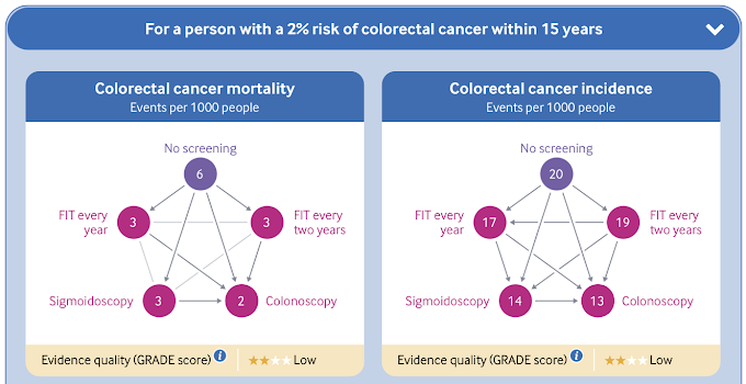
Dr. Toub ended his information-packed presentation with the suggestion that maybe we are thinking about CRC screening all wrong. In some well-resourced countries with national health plans, CRC screening is approached differently: rather than screening EVERYONE over a certain age, this BMJ guideline, published in 2019, suggests using a risk-based calculator to determine who should be screened. This suggests NOT screening anyone with <3% 15-year risk (based on this RISK CALCULATOR), which takes into account age, smoking and alcohol use, BMI, cancer hx among other things) to calculate that risk. IT also uses decision aids to help in determining screening decisons (see below):

What is the future of CRC screening?
It's AI, of course! Watch the last few minutes of Dr. Toub's talk for breaking news on AI in CRC screening. I think I'll leave it out of my summary today, as it is all still just starting and not really moved into standard practice.

For now,  remember these take home points: 1) CRC screening decreases deaths from CRC but does NOT improve all-cause mortality, 2) multiple screening modalities at multiple different intervals are considered equivalent, so use shared decision-making with your patients to meet their needs, and 3) use lots of different nudges to get your patients to follow-up with screening, but don't forget to mention the importance of CRC screening yourself!

Until next week. . .


Endometrial Cancer (Delic 11/8/2023)

A link to a recording of this presentation is available HERE

***
Many, many thanks to our speaker this week, Dr. Lejla Delic, a local gynecologist-oncologist, who works with our local oncologists and gynecologists to care for patients in our community with gyn cancers. Her presentation was excellent -- so good, in fact, that we we want her back for a Part 2, on ovarian cancer. Stay tuned. We will get her scheduled in the winter/spring.


In the meantime, my notes:

Gynecological cancers occur in the uterus (most common, aka endometrial cancer), cervix, fallopian tubes, ovaries, peritoneal cavity, vulva and vagina.

In the US, there are ~65,000 new cases of uterine cancer  per year --> 12,000 deaths
In the US, there are ~19,000 new cases of ovarian cancer per year--> 12,000 deaths

Unfortunately, mortality from uterine/endometrial cancer is on the rise

Endometrial cancer, the most common type of gyn cancers, has a lifetime prevalence of 3%. Women are average age 60 years old at the time of diagnosis and, because endometrial cancer typically presents with post-menopausal bleeding, 75% are diagnosed in the early stage (stage I and II). When it does metastasize, endometrial cancer typically spreads via local lymphatics to the pelvic nodes and then to the aortic lymph nodes. 

Unfortunately, black women in this country are dying at disproportionate rates of endometrial cancer and are diagnosed with more aggressive cancers, younger than white women. 

Risk factors for endometrial cancer:
  • Obesity (endogenous unopposed estrogen). In fact, every 5 points of BMI increases your risk of being diagnosed with endometrial cancer by 50%. 
  • Chronic anovulation (obesity, PCOS)
  • Nulliparity, infertility, early menarche, late menopause
  • Exogenous unopposed estrogen (e.g. HRT without progesterone)
  • Tamoxifen (2x risk of uterine cancer)
  • Hereditary: these present earlier than sporadic cancers, more typically in non-obese younger women (<50 years old), including genetic syndromes like Lynch Syndrome and MLH1, 2, 6, etc. 
Risk reduction for endometrial cancer:
  • OCPs (combined)
  • Weight loss
  • Hysterectomy (offered to women with Lynch syndrome after age 45 or when fertility is accomplished)
NEJM: Association with race and BMI and endometrial cancer


Historically, endometrial cancers have been divided in two groups: Type 1-- or non-aggressive, more excess estrogen type--  and Type 2, more aggressive, poorly differentiated, frequently metastasize (40% of women with Type 2 have +LN at time of diagnosis. Increasingly, however, new molecular characterization studies are changing the way we think about and treat endometrial cancer and are being integrated into the categorization. This is because prognosis is variable depending on these molecular characteristics.

90% of women with endometrial cancer present with post-menopausal (PMP) uterine bleeding. All cases of PMP bleeding should be investigated but it is important to note that only 9-14% of women with PMP bleeding have cancer.  Abnormal uterine bleeding (AUB) in premenopausal women may present more like "intermenstrual" bleeding.

Evaluation of AUB:
1) Pelvic ultrasound: in PMP women, >4mm endometrial stripe necessitates and EMB. Of note, endometrial stripe thickness in premenopausal women is totally useless. Also, in someone with repeated bleeds, even a thin stripe should not prevent you from getting an EMB.
2) Endometrial biopsy (EMB)

Of note, Type 2 Endometrial cancers, an EMB has a 25% false negative rate.
If the patient has recurrent bleeding, even if they have a normal EMB, refer for hysteroscopy and D&C

Treatment of endometrial cancer:

1) Surgery
All gyn cancers (except for cervical, trophoblastic and vaginal) need surgery to be staged.
Surgery includes hysterectomy, bilateral salpingectomy, and lymph node assessment (sential LN mapping).

Stage I: cancer confined to uterus
Stage II: cancer extends to cervix
Stage III: LN involvement/ovaries/tubes
Stage IV: distant mets (omentum, lungs)

Robotic/minimal invasive surgery has best outcomes (but NOT in cervical cancer). With robot, conversion to open only happens 3% of the time (whereas its 20% of the time with typical surgery). This is really important for patients with elevated BMI who have a much better/easier recovery with minimally invasive surgery and way less bad outcomes.

2) Immunotherapy may be indicated for Stage III or IV or recurrent endometrial cancer 
Historically, chemo was used (Carboplatin and paclitaxil) but with new molecular studies, immunotherapy is showing increasing promise.

Fertility preservation in young women with endometrial cancer is important to many women! 14% are pre-menopausal at diagnosis and may not be done building their families. Low Grade (1) and non-invasive cancers (as determined by MRI) can sometimes be treated temporarily with high dose progesterone (either oral Megace 80mg BID or Mirena IUD). Informed consent is important. About 50% respond, but 20-30% will either progress or return. Some women hate the side effects of high dose progesterone, which include increased appetite, weight gain, and blood clots. 

***

On a final note, Dr. Delic recommends TWO passes with your EMB pipelle when doing an EMB to ensure you get a good amount of tissue. And without a tenaculum whenever possible. 

Stay tuned for the spring for Part 2: Ovarian cancers

Big thank you to new-to-town-plastic surgeon, Dr. Melissa Mueller, for an excellent Grand Rounds presentation this week on Breast Reconstruction. I learned so much from her, and I look forward to working with her in our Sonoma County community. 

A recording of Dr. Mueller's excellent presentation is available HERE. If you care for women in Sonoma County, you should definitely watch this.

Here are my notes:

  • 1 in 8 women will develop breast cancer in their lifetime
    • 80% of women w/breast cancer have breast conserving therapy (i.e. lumpectomy)
    • of 20% of patients who undergo mastectomies, 40% get breast reconstruction, the majority implant based
    • in SoCo, less than the national average of patients get reconstruction, likely due to lack of access to modern breast reconstruction in the area
  • Breast reconstruction after cancer treatment is associated with improved self-image, decreased depression and anxiety, better body image, improved self esteem,  and overall improved emotional, social and sexual functioning
    • Some studies showing highest satisfaction with autologous reconstruction
  • The 1998 Women's Rights and Cancer Rights Act guaranteed reconstruction for women getting mastectomy (national)
  • California state law goes further and requires that ALL insurers (including Medi-Cal) must cover reconstructive surgeries (initial and subsequent) after breast cancer treatment, initial and subsequent implants (if patient needs an exchange)
    • includes mastectomy AND lumpectomy
    • includes contralateral breast symmetry procedures
  • Breast reconstruction may be a "silver lining" to getting cancer

Type of breast reconstruction after breast cancer depends on type of surgery.

After Lumpectomy (i.e. Breast Conserving Therapy or Partial Mastectomy)

All women in CA are eligible for oncoplastic reconstruction as well as contralateral breast symmetry

To be eligible for a lumpectomy (vs. mastectomy), the size of breast cancer must be small in relation to the size of breast. After the lump is removed, there will be a hole in the breast, which needs to be filled. This can be done in two different ways:

  • volume displacement: uses surrounding breast tissue to fill that hole--> that breast will be smaller and may require tissue rearrangement, reduction, lift/mastopexy
  • volume replacement: brings adjacent tissue outside the breast to restore the original breast size (e.g. back, side wall/bra fat, implants)
  • the contralateral breast will need to undergo symmetry procedure either immediately or delayed fashion 
    • this can happen at same time and/or after radiation
    • radiation causes collateral damage and decreased breast volume by 10-15%, tightening of the skin, nipple uplift
    • if reconstruction is happening before radiation, the breast that will be irradiated will be left slightly larger to allow for radiation changes
After Mastectomy, there are two categories of reconstruction
  • implant based: most commonly performed with tissue expander placed at time of mastectomy. Expander is inflated q1-2 weeks until desired size is achieved, then is replaced with implant (saline, silicone)
  • autologous: using patient's own tissue, make a breast mound with patient's own tissue, from abdomen/thigh/buttock/back (abdomen most common)


Reconstruction Timing 
post-mastectomy reconstruction also can happen immediately (at time of mastectomy) vs. delayed
    overall anesthesia time is less if doing delayed (in two parts)
    if done delayed, another advantage is that final pathology is received (margins) to know if radiation will be required

of note, breast skin cannot be expanded after radiation treatment, so if a patient wants an implant-based reconstruction, they need to have fully inflated before radiation tx
in advanced cancer, pts should focus on adjuvent therapy prior to reconstruction
but for patients who have the choice, more immediate reconstruction is associated with better outcomes

Implant-based reconstruction
under vs. over pectoralis mm
historically, implant has been placed under the muscle because the tissue makes implant less visible
however, recently prepectoral breast reconstruction is gaining in popularity-- less pain, no animation deformity (doesn't move when pectoralis moves)
fat grafting (i.e. liposuction) can help make the implant less visible in both cases

for some women, surgeons can place implant at time of mastectomy--> fewer total surgeries
    -have to have small breasts (A/B cup), want to stay same size, pts with nipple in correct place
    -mastectomy is stressor to breast skin, so heavy implants can create a second stressor to the skin which can compromise blood flow to the skin

Autologous reconstruction: 
create a breast mound using patient's own tissue (so you don't need an implant)
most common and most popular donor site is abdomen (deep inferior epigastric perforator flap)-- surgery similar to tummy tuck, kept alive by suturing to internal artery and vein in the chest
Fat grafting (ie liposuction--> injected with syringes after implant or autologous ( to disguise)


Counseling women for cosmetic results they may expect w/mastectomy
  • chest wall can be concave after surgery
  • lateral chest adiposity is not removed
  • dog ears (standing cone deformity)
BIA-ALCL
A word on very rare condition/complication from breast implants called Breast Implant Associated Anaplastic Large Cell Lymphoma
associated between textured devices-- both textured tissue expanders and implants (these are no longer used)
Incidence of association: 1/30K implants placed--> 949 total cases worldwide
On average 8-10 years after implant (at least 1 year)
Sudden fluid collection (collecting within a matter of days) or a new mass associated with the capsule around the implant
There has been a recall on all textured implants
FDA doesn't currently recommend removing the devices because it is unknown if removing them decreases lymphoma risk, but if patients feel uncomfortable having the device in their body, insurance will cover their replacement or removal
Ultrasound vs. MRI to evaluate, aspirate 50cc of fluid should be sent for pathology and immunochemistry


Diagnosis and Management of Osteoporosis (Hamann 7/23/2026)

 A recording of this presentation is available HERE .  *** Thanks so much to Dr. Kendal Hamann, SMGR Endocrinologist, for an outstanding Gra...