Showing posts with label dermatology. Show all posts

Neonatal and Infant Eruptions (Sugarman, 1/31/2024)

 A recording of this presentation can be found HERE

***

Many thanks to Dr. Jeff Sugarman, local dermatologist, who gave an excellent Grand Rounds presentation this week on Neonatal and Infant Eruptions: when to worry and when to reassure. See above for the link to watch it.

My notes this week come in the form of a dermatologic photo quiz for your testing pleasure. Answers are below the final photo and question.

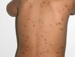
1. What is this neonatal rash categorized by fragile pustules that erode very quickly, leaving behind brown collarettes with post-inflammatory hyperpigmentation. It can be present at birth and last days to weeks. More common in neonates with more pigment. Benign.

Source: DermNetNZ.org
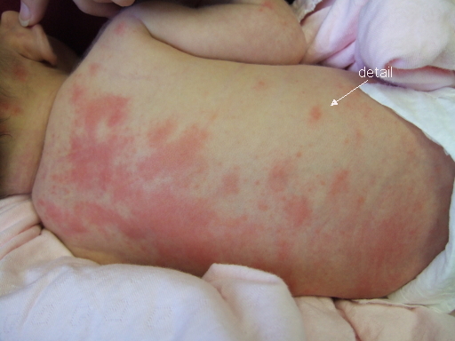
2. What is this benign newborn rash often caused by over-bundling newborns? Erythematous papules and pustules often occurring on covered portions of the skin. Totally benign.

Source: DermNetNZ.org

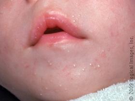
3. What is this benign rash of the neonatal period, often during the first 1-3 weeks of life, acneiform, often occurring on the face and upper chest and scalp. It has NO comedones and is the result of an inflammatory reaction. Treatment is either topical antifungal or  topical 1% hydrocortisone, though no treatment is also acceptable. Often mistaken for neonatal acne, which presents later AND always has comedones.

Source: DermNetNZ.org
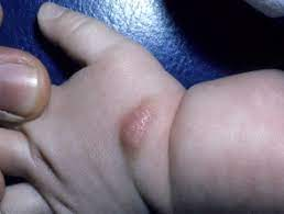
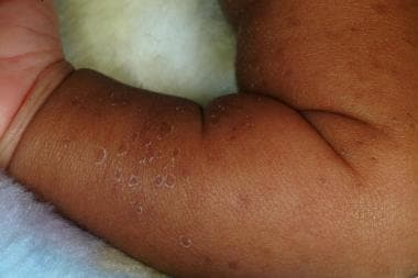
4. What is this benign self-resolving rash that is notable for pustules on feet and hands that can last for months, come and go in crops, and look a lot like scabies (but when scraped show no mites). This will resolve on its own but can take months to do so?

Source: DermNetNZ.org
5. What is this papule on the scalp, often solitary with a unique yellow color? The papule is self-resolving but can last years (5-10) before it disappears. Can sometimes occur in multiples and have extra-cutaneous involvement, with particular issues in the eye. In rare cases, multiple of these are associated with leukemia. 

Source: DermNetNZ.org
6) What is this condition characterized by peau d'orange (texture). Often missed by primary care clinicians. Usually solitary but can be present in sheets. Featured by pockets of histamine -- Darier's sign when stimulated (scratched or rubbed), can be accentuated. Can last for years. Activated by cold/hot/rubbing. 

Source: DermNetNZ.org
7) There are four variants of #6: 1) solitary (most common) 2) urticaria pigmentosa with peak onset first year of life 3) diffuse cutaneous mastocytosis and 4) most rarely mast cell leukemia. This is one of those variants, which features flushing, hives, pruritis, blisters and sometimes diarrhea (due to mast cells in the GI system). Treatment involves: antihistamines, oral cromolyn for GI symptoms, consider epi pen. If a high burden of disease, consider monitoring serial serum triptase.

Source: DermNetNZ.org
Of note, activators of mast cells include: alcohol, aspirin, narcotics (including codeine), some contrast agents, and hot/cold/sunlight. Consider limiting these triggers in patients with mastocytosis.

8) What is this eyebrow bump, often seen on the lateral brow, caused by a sequestration of ectodermal tissue during embryogenesis? It is usually present at birth but not noticed. It is rubbery,  not compressible, not tender. It is also not a true cyst. 

Source: DermNetNZ.org
Of note, you need to worry when the location suggests a CNS connection, which is always in the midline (25% of midline dermoids have a CNS connection). If a dermoid cyst is leaking clear fluid, this is likely spinal fluid and is BAD. It is good to remember that the neural tube closes in a series of discontinuous zippers, and there is no CNS connection in any other place than the "hot spots" where the zippers close. 

9) Do you have to worried about a CNS connection in this infant with a dermoid cyst?

Source: https://webeye.ophth.uiowa.edu/eyeforum/cases/115-dermoid-cyst.htm

Of note, dermoid cysts on the lateral brow are generally removed after a child's second birthday. They should be referred to pediatric plastics for the excision for best cosmetic effect. 

10) What vitamin deficiency -- often due to inadequate intake and appearing around the time a baby is weaning -- leads to  this psoriaform rash (well demarcated) on the face and in the diaper area? Hint: the name of the rash is acrodermatitis enteropathica.

Source: MDEdge
11) What is this uniquely pediatric form of this autoimmune disorder, often characterized by raccoon eyes (periorbital accentuation of erythema) and diagnosed by maternal serologies? Hint: photosensitive, 50% of cases have congenital heart block. Rare (10%) have hepatobiliary disease and even rarer (1%) thrombocytopenia.

Source: DermNetNZ

Source: DermNetNZ
12) Cool pearl!! Alternate treatment for umbilical granuloma. Aside from silver nitrate, a study showed that a single application of THIS common household item, covered with bandage tape can resolve this problem. What is the common household item?

13) What is this scary looking fixed erythematous rash (lasting 24+ hours) that often appears on the face, arms and legs. Child tends to be non-toxic appearing, and the trunk is spared. It resolves on its own. It is in the HSP spectrum, but has no cutaneous findings.
Source: DermNetNZ
14) Last but not least, what is this rash in a 6 week old baby that did not respond to topical steroid, topical anti-fungal, systemic antihistamines, or antibiotics?


And the bonus final: how do you treat it? Treatment of infantile scabies, even in children under 2 months and <15kg is the same as for children >2 months and >15kg (though the drug labeling does not promote this). Per Dr. Sugarman, you can confidently treat with either oral ivermectin OR topical permethrin safely and effectively in neonates.


********************
No cheating!!! Don't look down here until you have really thought about each of the photo questions above!!

Answers:

1) Transient Neonatal Pustular Melanosis

2) Miliaria Rubra

3) Neonatal cephalic pustulosis

4) Acropustulosis of infancy

5) Juvenile Xanthogranuloma

6) Mastocytosis

7) Urticaria pigmentosa

8) Dermoid Cyst

9) YES! This baby needs imaging (CT or MRI) to rule out encephalocele or mucocele prior to excision).

10) Zinc Deficiency: Treatment is rx zinc 3mg/kg/day

11) Neonatal Lupus Erythematosus

12) Table Salt!! A study of 17 infants with umbilical hernia found 100% resolution  (17/17) with a single application of table salt, occluded with medical bandage x 24 hours.

13) Acute hemorrhagic edema of infancy

14) Infant scabies, characterized by vesicles and pustules on the palms and the soles

Skin Cancer Reconstruction (Pourtaheri 4/13/2022)

Many thanks to Dr.  Navid Pourtaheri, new-to-our-community plastic surgeon, on Skin Cancer and Skin Cancer Reconstruction. This is a great summary of skin cancer findings and some specific indications and recommendations of when to involve plastic surgery. 

A recording is available HERE 

Skin cancer types: basal cell (BCC) squamous cell (SCC) keratoacanthoma, melanoma

Basal cell cancer (~4 million cases/year in US)

  • variable in appearance, "can look like anything"-- don't know what it is until you remove it
  • always slow growing (even high risk ones)
  • primarily occur on sun-exposed areas (hand, ears, face)
  • do sometimes spontaneous bleed or ulcerate
  • can be locally invasive
  • very rarely metastasize
Squamous cell cancer (SCC), 1.5 million cases/year in US
  • variable appearance, but more commonly dry and scaly, also can be rough/thickened, wart-like, can ulcerate and form open sores
  • more accelerated growth with local invasion than BCC (tend to progress more quickly)
  • lower lips more common with SCC 
  • can be locally invasive, can metastasize
  • curable if treated early
  • actinic keratoses (AK) is pre-squamous cell lesion
Keratoacanthoma, 275K cases/year in US
  • once considered subset of SCC
  • dome-shaped lesion with central keratin plug
  • almost always in sun exposed areas (face, ears, nose, hands)
  • quite rapidly growing (faster than SCC), can be locally destructive
  • unpredictable, can spontaneously regress
  • less likely to metastasize
Melanoma, 197K cases/year in US
  • most costly type of skin cancer
  • 25% found in existing moles, 75% spontaneously occur on normal looking skin
  • 50% are melanoma in situ>> high cure rate (90-100%)
  • can be found ANYWHERE
  • most common form is superficial spreading
  • when goes deeper, called lentigo maligna
  • 10-15% of cases are nodular melanoma, most aggressive, tends to be found invading
  • remember ABCDE (see below) with special attention to "E" (evolving-- that is a changing mole)
  • Breslow thickness correlates with 5-year survival


Treatment options for skin cancer
  • Moh's surgery 
    • most commonly used in BCC and SCC (often contraindicated for melanoma)
    • high risk areas (H zone)
    • cosmetically sensitive areas, over joint surfaces
    • decreases amount of tissue excised, maintaining the same cure rate
  • Excision with margins and simple closure also acceptable
  • Curettage (EDC)
    • scrape the abnormal tissue, then burn it
  • Cryotherapy more common in older patients, people who don't want a procedure, on blood thinners, large number (e.g. on face)
  • Melanoma best excised w/margins, possible lymph node biopsy (plastic surgery, general surgery)
    • sentinel node biopsy not indicated for melanoma in situ
When to refer to plastic surgeon?
  • skin cancer not indicated for Mohs
  • post dermatology resection, cannot close
  • melanoma
Biopsy options
  • shave: get full epidermis but only part of dermis (this is good because it won't form scar)
    • always inappropriate for c/f melanoma because need full thickness
  • punch: takes full thickness of skin
  • incisional biopsy: get piece of abnormal with normal adjacent, done with scalpel
  • excision: preferred for lesion is <1cm (cut the whole thing out with margin, 1mm of normal tissue is acceptable margin 
  • FNA not used for skin, but for clinically positive nodes
  • Sentinel lymph node biopsy (SLNB) after melanoma diagnosis, after lymphoscintigraphy (radioactive dye that drains to the lymph node)
Imaging
  • Always needed in >Stage 3 melanoma, definitely NOT indicated in stage 1 (stage 2, use your judgement, may be indicated)
  • Most common: PET CT for assessing for metastases
  • Chest CT also good modality b/c melanoma so often goes to lungs (chest xray not sufficient)

What a Pain! Tales of Adult Arthritides (Ramirez, 3/31/2021)

Thank you to Dr. Vanessa Ramirez for her review of two very important inflammatory arthritides at Grand Rounds this week: psoriatic arthritis and gout.  I gleaned so many important pearls from her presentation. For those of you interested in seeing the full recording, it is available here. My summary notes are below:

Inflammatory arthritides

  • infectious (septic)
  • crystal induced (gout, pseudogout)
  • immune related (RA, SLE, psoriatic arthritis, dermatomyositis, Sjogren's)
  • reactive
Psoriatic arthritis (PsA) 

  • PsA affects about 20% of people with psoriasis 
    • skin changes can precede arthritis sx for years-- even up to 12 years
    • skin changes and PsA flares are not necessarily temporally related, nor is disease severity necessarily correlated
  • typically asymmetric arthritis (in one, several or multiple joints), sacroiliitis (30-78%) 
  • historic definition of PsA: serological negative (i.e. negative rheumatoid factor) polyarticular arthritis in someone with psoriasis skin manifestations
  • classification Criteria for Psoriatic Arthritis (CASPAR) (2006) may be helpful in making this clinical diagnosis (see image below)
  • early identification and treatment prevents joint destruction
  • anti-CCP may be elevated (usually mild) in 12% of patients with PsA
    • more likely in higher numbers of involved joints
  • Risk factors for PsA
    • scalp psoriasis 4x risk, intergluteal/perianal 2.3x risk, nail involvement
    • earlier age at dx with psoriasis, >3 body sites affected, family hx of PsA (first degree)
  • ESR is superior to CRP as a marker of damage progression and mortality
  • Treatment for PSA includes lifestyle modification (diet, smoking cessation, exercise), followed by symptomatic treatments (NSAID, steroids, injections) and then TNF alphas
    • see images below from the 2018 ACR Guidelines
    • also see AAFP image, which includes cost of these treatments

2018 ACR Guidelines for Treatment of PsA
https://www.rheumatology.org/Portals/0/Files/PsA-Guideline-2018.pdf

AAFP Psoriasis (Am Family Physician 2013)
https://www.aafp.org/afp/2013/0501/p626.html

Gout 
  • Gout is caused by deposition of monosodium urate crystals in the joint space, periarticular structures and soft tissues
    • associated with obesity, htn, hyperlipidemia, DM, CKD, heart failure, thiazide diuretic 
  • ACR online tool Clinical prediction (see image)
  • https://www.aafp.org/afp/2020/1101/p533.html


  • Treatment Acute
    • 2020 ACR Guidelines for Treatment of Gout Flare
      • Naproxen 500 mg BID OR indomethacin 50 mg TID, ibuprofen 800mg TID
      • Colchicine (low dose) 1.2mg PO, then 0.6mg 1 hour later, then BID until flare resolves
      • Oral prednisone 0.5mg/kg (5-10 days full dose then stop OR 2-5 days full dose and then taper over 7-10 days)
  • Treatment Chronic
    • all patients with tophi, radiographic evidence or damage or 2+ flares/year
    • goal is symptom relief AND maintenance of urate levels (<6)
      • we should be titrating allopurinol based on checking uric acid levels
    • Allopurinol is treatment of choice, lower doses preferred to start
    • if on thiazide for BP, switch to losartan


Dermatologic Emergencies and their Mimics (Sugarman, 9/30/2020)

 Thanks to Dr. Jeff Sugarman for an excellent Grand Rounds this week on Dermatologic Emergencies and their Mimics. Dr. Sugarman's presentations are always replete with photos ("A picture is worth a thousand words" for sure) and probing questions, so this post will be filled with the same. Answers can be found at the very end of the post in the COMMENTS section. Don't cheat; take the quiz and use the HINTS not only to guide you to your answers, but also to enhance your understanding of the condition. 

First, when should you worry about possible dermatologic emergencies?

  • Age (newborn and young infants)
  • High fever, toxicity
  • Morphology: particularly blistering, mucosal involvement, hemorrhage
  • Specific medications: anticonvulsants, antibiotics, NSAIDs
Remember the presentation was on dermatologic emergencies and their mimics. This summary/quiz contains both derm emergencies and benign derm conditions that look pretty similar, so keep serious and not serious things on your differential. 

1) What is this rash?

Hint #1: it's really common (especially in children and people with atopy)
Hint #2: morphology includes wheals, annular, dusky centers
Hint #3: time course is VERY helpful: lesions tend to self resolve in hours, disappear and reappear in different locations
Hint #4: triggers include allergy, autoimmunity, drugs (9%), URI (40%), and idiopathic (50%)
Hint #4: Treatment: non-sedating antihistamine (fexofenadine, cetirizine) in day, sedating antihistamine at night (hydroxyzine, diphenhydramine). 
Hint #5: Prednisone is NOT rx of choice-- it works really well, and then the rash will come right back as soon as it's stopped.

2) What is this rash?
Hint #1: Looks a lot like the first rash but is different.
Hint#2: Rash morphology includes target lesions with 3 zones: dusky center, pale edematous ring, peripheral erythematous margin
Hint #3: lesions are discrete, they do NOT coalesce
Hint #4: usually pts have no systemic symptoms

3) What is this rash?

Hint#1: Presents as dusky urticaria PLUS edema, +/- fever, malaise and arthritis (7-21 days after exposure)
Hint #2: Lesions last longer than true urticaria
Hint #3: This is a type III hypersensitivity reaction (immune complexes)
Hint #4: Triggers include meds (cefaclor, PCN, anti-cancer, anti-depressants, anticonvulsants, htn meds, anti-inflammatory meds), biologic agents (rituximab, infliximab, efalizumab), infections (strep, HBC, HCV)

4) What is this rash?


Hint #1: This is a form of leukocytoclastic vasculitis in children age <2 years old
Hint #2: Presents as purpuric edematous plaques with target-like pattern, often described as "cockade or rosette"
Hint #3: This includes dramatic skin findings, but children paradoxically are not really toxic
Hint #4: Rash tends to spare the trunk
Hint #5: Lesions resolve spontaneously in 1-3 weeks
Hint #6: No labs or treatment needed.

5) What is this rash?

Hint#1: This rash may accompany pneumonia by this same organism
Hint #2: Tends to be mucosal predominant (94% oral, 82% ocular, 63% GU) and is mucosal alone in 34% of cases
Hint #3: Mean age is 12 years old
Hint#4: Most patients (81%) have no long term sequelae
Hint #5: I never heard of this before this lecture by Dr. Sugarman

6) What is this rash?


Hint#1: Severe life-threatening mucocutaneous disease involving systemic signs: fever, respiratory symptoms
Hint #2: It's a clinical syndrome, there is no definitive diagnostic test
Hint #3: Always involves at least 2 mucous membranes (mouth, eyes, urethra)
Hint #4: Causes in kids include meds (antibiotics, antiepileptics, chemotherapy), as well as HSV, mycoplasma and some undetermined causes

7) What is this rash?

Hint #1: begins as localized often occult infection (can be in the nasopharynx, perioral, conjunctiva, umbilicus, paronychia, urine, middle ear)
Hint #2: Progresses to generalized erythema and skin fragility
Hint #3: Empiric treatment is anti-staph antibiotics (cover for MRSA)
Hint #4: Peeling is NOT full thickness

8) What is this rash?
Hint #1: Most common cause of nonsexually related acute genital ulcers (NRAGU)
Hint #2: Ulcers are painful, well demarcated, shallow erosions on a clean fibrinous base
Hint #3: Self-limiting condition, usually resolving spontaneously within 2-6 weeks

9) What is this rash?

Hint #1: thick crusts, thick walled pustules are common
Hint #2: facial, periorbital involvement common 
Hint #3: fever and pain are common
Hint #4: You should culture this
Hint #5: Keflex and mid-potency steroid for body (TAC) and low potency steroid (2.5% hydrocortisone) are both indicated
Hint #6: Bleach baths (1/2 cup in full bath, 1/4 cup in 1/2 bath) may also be indicated

10) What is this rash?

Hint #1: People with eczema are particularly vulnerable to this condition due to their disruption of epidermal barrier
Hint #2: Fever, malaise, and lymphadenopathy may be present
Hint#3: This is PAINful
Hint #4: Morphology includes "monomorphous punched out erosions" (especially if you look at the periphery of this rash)
Hint #5: Lesions favor areas of active dermatitis, particularly head, neck and trunk
Hint #4: There is often a delay in diagnosis of this condition
Hint#5: Viral culture/PCR will give you the answer
Hint #6: Prompt high dose acyclovir is treatment of choice (PO for mild, IV for mod/severe)

Neonatal Dermatology (Sugarman, 2/12/2020)

Muchas Gracias to Dr. Jeff Sugarman for a solid tour through neonatal dermatology this week.


My biggest take home: IF/WHEN you see a hemangioma in a newborn on the face, refer them IMMEDIATELY to dermatology. That day, says Dr. Sugarman. Medical treatment is time sensitive, and you could be saving them future surgery.

Just for kicks, I am going to gift you with a photo quiz rather than a written summary.  Good luck (answers at the VERY bottom of this post):

Identify the following benign non-infectious lesions in neonates:
Image result for erythema toxicumImage result for sebaceous hyperplasia neonateImage result for milia neonateImage result for neonatal cephalic pustulosisImage result for transient neonatal pustular melanosisImage result for acropustulosis of infancy


And now for infectious causes of newborn eruptions:
Image result for impetigo neonatorumImage result for scabies neonate

Image result for congenital candidiasis
Image result for neonatal hsv


And finally, some really common markings and some zebras:
Image result for nevus simplex
Image result for scalp aplasia cutisImage result for hemangioma face neonate
Image result for nevus sebaceous neonate Image result for neonatal lupus

Don't cheat and look below until you have taken this quiz yourself!!! 
 ___________________________________________________________________
ANSWERS:
First row: benign lesions
·         Erythema toxicum
·         Sebaceous hyperplasia
·         Milia
·         Neonatal cephalic pustulosis
·         Transient neonatal pustular melanosis
·         Acropustolisis of infancy
Second row: infectious  lesions
·         Impetigo Neonatorum
·         Scabies
·         Congenital candidiasis
·         Neonatal HSV
Third row  
·         Nevus simplex
·         Hemangioma
·         Scalp aplasia cutis
·         Nevus sebaceous
·         Neonatal lupus

Diagnosis and Management of Osteoporosis (Hamann 7/23/2026)

 A recording of this presentation is available HERE .  *** Thanks so much to Dr. Kendal Hamann, SMGR Endocrinologist, for an outstanding Gra...