Showing posts with label surgery. Show all posts

Spine Surgery for the PCP (Athanassious, 7/15/26)

A recording of this presentation is available HERE.

Many thanks to Dr. Christian Athanassious, orthopedic spine surgeon, who gave a really great Grand Rounds Treatment of the Spine for the PCP. The recording is worth watching! For those who prefer the written word. . .

Key learning points:
-The cervical and lumbar spine contains mobile segments>> most likely to develop problems (in contrast to thoracic spine)
-Cervical DJD can often radiate to the scapula
-Be sure to give high enough doses of gabapentin (e.g. 300mg QHS) for acute back pain, helping pts sleep improves pain
-Myelopathy with acute change in function (e.g. ability to hold a cup, balance) needs urgent eval
-If patient has >6 months pain with stenosis on MRI, they are unlikely to get better and should be referred to spine surgeon
-Spinal precautions: no twisting, bending or lifting > 10 pounds (fusion x  6 weeks, then can increase weight to 25 pounds x 3 months, laminectomy x 6 weeks)

Neck Pain
-Radiculopathy: compression of cervical nerve roots: deep aching, electrical feeling, stabbing, numbness/tingling (must be differentiated from carpal tunnel syndrome)
-DJD means "less hydration in the disk level" (doesn't mean there is compression nerve or pain generator)-- most adults will have 1-2 levels of DJD on imaging, must always correlate with history and physical exam findings
-Most common cervical problem is C5/C6:  neuroforaminal narrowing will affect the C6 nerve root (weak biceps, wrist extension, deltoid weak)
-Second most common: C6/C7: numbness and tingling over forearm, 2nd and 3rd digit (radial), wrist flexion weakness, triceps weakness
-if a cervical problem > 6 months, should refer to spine surgery

-Myelopathy: can present as a step-like decline (from one day to next), more urgent than radiculopathy. Pain doesn't follow dermatomal distribution because whole spinal cord is being compressed
-while nerve roots can recover (even after 1-2 years of injury), spinal cord injury does not recover as well
-surgical emergency if acute change in function
-"I was walking with a cane independently, and now I feel like I am drunk, I cannot control my feet, I do not know where my arms/legs are in space">> needs cervical and thoracic MRI and spine surgery consultation, may need ED visit
-sudden change in handwriting, in ability to hold a cup
-bowel and bladder dysfunction are rare (need major compression)

Lumbar Pain
Less urgent low back pain: sciatica/back pain
More urgent: cauda equina
L1-L5/Sacrum (6 vertebrae)
Discs: annulus fibrosis (outer fibrous layer), nucleus pulposus (gelatinous)>> pain from annulus being torn, another part is nerve roots being compressed or the pulp putting pressure on the nerves  caustic to nerve root, can cause pain). Acute annular tear, release of NT and tear can cause acute pain>> PT, back precautions, NSAIDSs vs. oral steroids

Non-operative management of acute lumbar disc herniation
-NSAID
-Physical therapy
-gabapentin/lyrica: make sure to dose gabapentin high enough
-oral steroid
-referral to pain mgt for epidural steroid injection
-brace (for fracture, first line for fracture tx unless severe compression of nerve root, patient with a body habitus that doesn't allow, or emphysema dx cannot tolerate) >>vertebroplasty/kyphoplasty: spine surgeon, IR, pain mgt surgeon (okay to call spine surgery if we have questions about what to do)

Ddx lumbar Pain: arthritis, stenosis, DJD (if bad enough), facet disease

Back symptoms:
-axial back pain: doesn't radiate (stenosis, arthritis in facet, severe DJD leading to bone on bone)
-radicular: irritates the nerve root, radiates down legs or out to arms, or to scapula (if thoracic, can radiate mid back toward ribs)
-loss of balance (spinal cord compression)
-loss of dexterity (spinal cord compression)
-weakness (can happen with nerve root or spinal cord compression)

***
Neck surgery:
-Most common: Anterior cervical discectomy and fusion (ACDF), less common disc arthroplasty (if disease limited to 1-2 levels)
-Disc removed, spinal cord and facet decompressed>> spacer placed. If fusion, plate placed over the top (if just arthroplasty, no plate placed)
-Disc arthroplasty: not indicated for >2 disc levels, facet disease often a problem. Usually indicated only in young patient with little facet disease, short hx of axial neck pain, acute issue 2/2 fall or accident (disc replacement). Many will eventually need a fusion

Normal ACDF recovery: 
    -some degree of difficulty swallowing (no need for swallow study), have to chew longer, cut smaller. Pay attention when swallowing. Can last weeks to 1-2 months, almost always resolves
    -pain in front of neck
    -mild swelling in front of neck>> make sure breathing okay, can speak, eat and drink. Can consider TXA post-op to prevent seeping
    -mid-scapula pain: spacers cause stretch on facets and capsule and can cause posterior neck that radiates to scapula
    -strength doesn't come back right away, can continue to see recovery up too 2 years post-op
    -neck braces after fusion (1 level 4 weeks, 2 levels 8 weeks, 3 level 12 weeks in brace)

Abnormal ACDF recovery:
    -C5 radiculopathy (difficulty lifting shoulder) common after severe spinal stenosis due to posterior drift, not permanent, give brace so arm not hanging, will resolve over time
    -redness/infection of surgical site (if no fever, can rx cephalexin; if fever, may need admission for IV abx, MRI to ensure infection is superficial)
    -any NEW dense weakness>> get MRI
    -Horner syndrome: symptomatic treatment, lubricating drops for eyes
surgical decompression (microdiscectomy)

Low Back Surgery
    -microdiscectomy most common
    -laminectomy (full removal) vs. laminotomy
    -#1 indication for spinal fusion is mobile spondylolisthesis (pt flexes and extends and has >3mm motion between flexion/extension). If spinal stenosis and symptoms correlate, not improving 3-6 months, intolerable of pain
    -Why fuse?
        -DJD with stenosis
    -Adjacent level disease
        -degenerative scoliosis

Access lumbar spine from the 1) front (anterior retroperitoneal approach): incision in midline of stomach, go around peritoneum, fuse in retroperitoneal. 2) lateral anterior retroperitoneal approach, 3)  posterior fusion

ALIF: anterior lumbar interbody fusion. Very stable, access via vascular surgery (higher risk of bleeding), powerful way to treat patients IF treating L5/S1 (cannot be accessed frmo the side). Downsides: post-op bleeding, post-op infection (superficial vs. deep (abnormal labs, fever/vital sign instability), DVT/PE (increased due to vessel manipulation), sympathetic chain compromise 

PLIF: posterior lumbar interbody fusion. Less effective, more problematic. Have to take down a lot of paraspinal musculature, removal of bone and ligament from posterior canal. Cannot correct deformity as well as ALIF. 

EXLIF: minimally invasive procedure, eXtreme lateral interbody fusion. Much smaller incision (compared to PLIF). Can use large cage. large graft to increase stability. L5-S1 cannot be accessed due to the anatomy (pelvis in the way). Can get anterior thigh numbness/hip flexion weakness (psoas mm)


What requires urgent spine surgery attention?
-Myelopathy: quick change in function, send to ED, call spine surgeon
-Severe radiculopathy with weakness
-If symptoms > 6 months, doesn't need ED eval but should refer to spine surgery (especially if MRI finds stenosis)
-Cauda equina: severe excruciating back pain, weakness on exam, numbness ins addle, bowel/bladder incontinence. Usually caused by acute event (should be treated within 48 hours, bowel and bladder function may not return)

Perianal Disease: Not Everything down there is a Hemorrhoid (Cortez, 11/20/2024)

 A recording of this presentation is available HERE

Many thanks to Dr. Allen Cortez for an excellent presentation on perianal disease. I learned a lot and was really impressed by how much Dr. Cortez, a long-time local general surgeon, knows and cares about a region of the body that many people are pretty uncomfortable talking about. I recommend you watch his presentation, the link above. 

My notes:

Hemorrhoids are extremely common -- 5-10% of the population, >2.2 million people per year with over 2 million prescriptions per year that add up to over $43 million in healthcare costs. But many patients are being treated for hemorrhoids when there may be other things going on down there, including: fissures, fistula, abscess, pruritis, and rectal prolapse. 

Dr. Cortez reminded us to go back to the basics: 1) listen to the patient (e.g. hemorrhoids generally don't hurt, so if the patient is complaining of pain, broaden your ddx) and 2) examine the patient.

Fiber

Encourage fiber! All our patients need more fiber. And fiber isn't good for just the perianal region. Fiber decreases risk of cardiovascular disease, decreases risk for colon cancer. "The best fiber out there is the one you'll take". Really, the only downside is increased flatulence.

Fiber options abound, including: psyllium husk, benefiber (can sprinkle on yogurt), fiber capsules or gummies (e.g. Kirkland brand, 2-4 gummies 2x per day, with LOTS of water).

Once you start a fiber supplement, don't make any changes for 5 days.

Miralax is good too, but it shouldn't replace fiber. Use miralax PRN for constipation. 

Dr. Cortez reminded us that the increased pressure of diarrhea can also contribute to hemorrhoids. The goal is ONE nice big healthy bowel movement per day, no more than 4-5 minutes sitting on the pot. It is better to return 3 or more times than sit for prolonged period of time on the toilet.

When you are going to examine a patient for perianal complaints: put them in L lateral decubitus (better to see than lithotomy). Look externally for tags, fissures/openings, thickened skin, ulcerations, masses. If you see a fissure stop there (you can make it worse). Use anoscopy and a digital exam to check for tone, masses, blood, etc. 

Dr. Cortez is not a fan of donuts for any perianal condition. In his eloquent words: "Gravity tries to push your liver right out your butt"

Hemorrhoid Treatment 

1) Banding (in office), no prep, effective, can be done several times, no downtime, no severe pain

2) Hemorrhoidectomy (surgical) is always a last resort, very pain ful but most effective. Stapled hemorrhoidectomies are not superior.

Thrombosed hemorrhoids, which present as big purple extremely painful lumps should be unroofed in first 2-3 days for pain control (in ED or office). If it has been > 5 days, healing is equivalent and intervention is not indicated.

Fissures

Perianal fissures, which are tears in the anoderm exposing the sphincter muscles, are extremely painful. Patients may describe symptoms as "crapping out glass" or "jamming a knife in my butt". 73% present midline posteriorly. If you visualize a lateral fissure, that patient needs a work-up, including testing for Crohn's, HIV, syphillis, TB and more. 


Acute fissures (< 6 months) can be treated with fiber, fiber and fiber, as well as hydration and sitz baths. This takes time! Topicals can sooth and manage symptoms, including topical nitroglycerine. Dr. Cortez's preferred topical is compounded diltiazem/nifedipine cream (locally can get compounded at Dollar Drug). Can be rx'd TID.

Chronic fissures (>6 months) require treatment with chemo-denervation, including Botox, which is effective and stops spasm. Some people need surgical intervention: with sphincterotomy or anocutaneous flap.

Perianal abscess

Perianal abscesses can also be extremely painful. 30-70% of abscesses have an accompanying fistula. 40-50% will develop a fistula over time.

 Treatment of perianal abscess is I&D, and usual management with packing is not effective because the skin often heals over before the abscess heals. Instead make a BIG incision (this may require sedation-- need to happen in ED?) and may need an initial packing but then should probably not be packed . Perianal abscesses can occur in several locations: ischiorectal, intersphincteric, perianal and supralevator. No antibiotics are indicated once drainage (i.e. source control) is achieved. Many fistulas will heal themselves and not all perianal abscesses need CT imaging. But if you are concerned, send to surgeon for further assessment. 

Malignancy

Finally, malignancies can present in the perianal region and the only way to diagnose them is to look for them. These can include squamous cell carcinomas and melanomas. See images below for some examples. 


Perioperative Evaluation (Schneider - 10/4/23)

Sorry, there is no recording available for this session. 

***

Thank you to our very own Dr. Dave Schneider for an excellent presentation on Perioperative Evaluation. Unfortunately, I forgot to hit "record" on the zoom meeting, so we do not have a recorded version of his presentation. Sorry about that!

My notes:

First off, "clearance for surgery" is not our job!  Our job is to assess each patient's perioperative risks and optimize and manage those risk factors as they head into surgery. Do note that there are gender and race disparities in those who receive surgical interventions (BIPOC patients receive fewer PCI interventions, fewer orthoplasties and have increased mortality when undergoing these procedures).

The new-ish term for perioperative cardiovascular complications is Myocardial Injury after Non-Cardiac Surgery (aka MINS). 5-19% of surgical patients will experience MINS, 84% will be asymptomatic. MINS is associated with increased morbidity and mortality.

The American College of Cardiology (ACC) last updated their Guidelines for Perioperative Cardiovascular Evaluation and Management for patients undergoing non-cardiac surgery in 2014. These old guidelines are available here. The flow diagram is a doozy and the recommendations are confusing: 

In classic Schneider-fashion, Dr. Schneider invented an acronym to summarize their 2014 recommendations. It is called E-A-R-L-I

E: Emergent: if a surgery is emergent--> take patient to the OR and deal with the negative outcomes later

A: ACS: if the patient has s/sx of ACS, manage per guidelines

R: Risk assess --> use any of several tools (e.g. RCRI, NSQUIP calculator, MICA calculator, see below for more info)

L: Limitation of function --> if patient unable to  perform at 4 METs using the Duke Activity Scale, optimize their functional status before proceeding to surgery

I: Impact on decision? --> Yes or No? If the cardiac stress test outcome will change how/when you will proceed with surgery, then go ahead with a stress test. If not, then proceed to the OR.

The European Society of Cardiology (ESC) updated their guidelines on perioperative management more recently in 2022. And their guidelines are much more simple than those of the ACC. They are summarized here by the ACC. In essence, they say: 

Is the surgery. . .

Emergent? -->  Proceed to surgery without delay, cardiac testing is not feasible

Urgent? --> Proceed to surgery without unnecessary delay (using a multidisciplinary team to determine about individualized cardiac testing)

Time-Sensitive? --> Do the surgery ASAP

Their guidelines are summarized in this lovely flow charts. Don't you just love flow charts?


***

Should you check a Hemoglobin and Renal function in all patients pre-operatively? The answer is no, but your should check in intermediate and high risk patients. 

Don't forget, for everyone, advise smoking cessation!

There are two risks to consider in evaluating patients: 1) the risk of the surgery itself (e.g. highest risk includes intra-thoracic, vascular) and 2) the risk of the patient

  • If the surgery is low risk, no CV assessment needs to be made
  • If the surgery is intermediate risk, and the patient is either >65 or with CV risk factors, get an EKG and check functional capacity
  • If the surgery is HIGH risk, consider EKG and biomarkers** for patients older than 45, definitely get them for patients >65 or with CV risk factors. If the patient has known CVD, get a cardiology consultation and make a multidisciplinary decision. 
**Note: Biomarkers referred to above include BNP and/or Cardiac Troponin (also in table above). They have been shown to predict MI. Either one is predictive and do not change outcomes. 

There are several risk calculators to help you determine your patient's  risk level:
1) Revised Cardiac Risk Index (RCRI), which Dr. Schneider shortens to DRC4 (diabetes, risky surgery, CAD, CHF, CVD, Cr>2)
Each calculator is slightly different and variably useful depending on the patient in front of you. All three of these have been validated and you should get familiar with all of them.

Okay, now for a few pearls:
  • There is NO benefit to coronary revascularization before surgery.
  • Labs and other tests should only be done pre-operatively if you were going to do them anyway
  • Coag testing is usually unnecessary unless patient is on warfarin. Family Hx and PMH are just as predictive of bleeding (e.g. if patient has history of prior bleed, or family member has bleeding problem, patient has higher risk of bleed)
  • Only get a pre-op EKG if the patient has known CVD, CV risk factors >65 and they are having an intermediate/high risk surgery
  • TTE only needed if patient has known valvular lesion and no TTE in the last year. You may consider if new onset dyspnea or change in status of their HF
  • Pre-op CXR is NOT recommended (Choosing Wisely, ACR 2017)
What about medications?
Statins: if a patient is on a statin, continue it (okay to miss a few days due to NPO, etc.). Perioperative initiation is reasonable if someone is getting vascular surgery

Beta blockers: if patient is already on BB, continue them perioperatively (perioperative withdrawal has 4x increased mortality). You may consider decreasing BB dose due to risk of hypotension after surgery. You can consider starting a BB at least one week (up to 28 days) prior to cardiac surgery if high risk patient and high risk surgery. 

Other anti-hypertensives: post-op hypotension is a common problem. Consider holding all BP meds on day of surgery, add them back slowly post-op, ?one at a time

ASA: it is okay to go to the OR on aspirin. Also okay to stop ASA in high bleeding risk patients (e.g. those on DOAC or warfarin as well). Continuing ASA has been shown to be cardioprotective: decreased MI by 56% and decreased composite CV outcomes. There is a non-significant increased bleeding risk if you continue ASA.

What about patients with recent drug eluting stents (DES)? Delay elective surgery for up to 6 months if possible so as not to interrupt DAPT. 


Big thank you to new-to-town-plastic surgeon, Dr. Melissa Mueller, for an excellent Grand Rounds presentation this week on Breast Reconstruction. I learned so much from her, and I look forward to working with her in our Sonoma County community. 

A recording of Dr. Mueller's excellent presentation is available HERE. If you care for women in Sonoma County, you should definitely watch this.

Here are my notes:

  • 1 in 8 women will develop breast cancer in their lifetime
    • 80% of women w/breast cancer have breast conserving therapy (i.e. lumpectomy)
    • of 20% of patients who undergo mastectomies, 40% get breast reconstruction, the majority implant based
    • in SoCo, less than the national average of patients get reconstruction, likely due to lack of access to modern breast reconstruction in the area
  • Breast reconstruction after cancer treatment is associated with improved self-image, decreased depression and anxiety, better body image, improved self esteem,  and overall improved emotional, social and sexual functioning
    • Some studies showing highest satisfaction with autologous reconstruction
  • The 1998 Women's Rights and Cancer Rights Act guaranteed reconstruction for women getting mastectomy (national)
  • California state law goes further and requires that ALL insurers (including Medi-Cal) must cover reconstructive surgeries (initial and subsequent) after breast cancer treatment, initial and subsequent implants (if patient needs an exchange)
    • includes mastectomy AND lumpectomy
    • includes contralateral breast symmetry procedures
  • Breast reconstruction may be a "silver lining" to getting cancer

Type of breast reconstruction after breast cancer depends on type of surgery.

After Lumpectomy (i.e. Breast Conserving Therapy or Partial Mastectomy)

All women in CA are eligible for oncoplastic reconstruction as well as contralateral breast symmetry

To be eligible for a lumpectomy (vs. mastectomy), the size of breast cancer must be small in relation to the size of breast. After the lump is removed, there will be a hole in the breast, which needs to be filled. This can be done in two different ways:

  • volume displacement: uses surrounding breast tissue to fill that hole--> that breast will be smaller and may require tissue rearrangement, reduction, lift/mastopexy
  • volume replacement: brings adjacent tissue outside the breast to restore the original breast size (e.g. back, side wall/bra fat, implants)
  • the contralateral breast will need to undergo symmetry procedure either immediately or delayed fashion 
    • this can happen at same time and/or after radiation
    • radiation causes collateral damage and decreased breast volume by 10-15%, tightening of the skin, nipple uplift
    • if reconstruction is happening before radiation, the breast that will be irradiated will be left slightly larger to allow for radiation changes
After Mastectomy, there are two categories of reconstruction
  • implant based: most commonly performed with tissue expander placed at time of mastectomy. Expander is inflated q1-2 weeks until desired size is achieved, then is replaced with implant (saline, silicone)
  • autologous: using patient's own tissue, make a breast mound with patient's own tissue, from abdomen/thigh/buttock/back (abdomen most common)


Reconstruction Timing 
post-mastectomy reconstruction also can happen immediately (at time of mastectomy) vs. delayed
    overall anesthesia time is less if doing delayed (in two parts)
    if done delayed, another advantage is that final pathology is received (margins) to know if radiation will be required

of note, breast skin cannot be expanded after radiation treatment, so if a patient wants an implant-based reconstruction, they need to have fully inflated before radiation tx
in advanced cancer, pts should focus on adjuvent therapy prior to reconstruction
but for patients who have the choice, more immediate reconstruction is associated with better outcomes

Implant-based reconstruction
under vs. over pectoralis mm
historically, implant has been placed under the muscle because the tissue makes implant less visible
however, recently prepectoral breast reconstruction is gaining in popularity-- less pain, no animation deformity (doesn't move when pectoralis moves)
fat grafting (i.e. liposuction) can help make the implant less visible in both cases

for some women, surgeons can place implant at time of mastectomy--> fewer total surgeries
    -have to have small breasts (A/B cup), want to stay same size, pts with nipple in correct place
    -mastectomy is stressor to breast skin, so heavy implants can create a second stressor to the skin which can compromise blood flow to the skin

Autologous reconstruction: 
create a breast mound using patient's own tissue (so you don't need an implant)
most common and most popular donor site is abdomen (deep inferior epigastric perforator flap)-- surgery similar to tummy tuck, kept alive by suturing to internal artery and vein in the chest
Fat grafting (ie liposuction--> injected with syringes after implant or autologous ( to disguise)


Counseling women for cosmetic results they may expect w/mastectomy
  • chest wall can be concave after surgery
  • lateral chest adiposity is not removed
  • dog ears (standing cone deformity)
BIA-ALCL
A word on very rare condition/complication from breast implants called Breast Implant Associated Anaplastic Large Cell Lymphoma
associated between textured devices-- both textured tissue expanders and implants (these are no longer used)
Incidence of association: 1/30K implants placed--> 949 total cases worldwide
On average 8-10 years after implant (at least 1 year)
Sudden fluid collection (collecting within a matter of days) or a new mass associated with the capsule around the implant
There has been a recall on all textured implants
FDA doesn't currently recommend removing the devices because it is unknown if removing them decreases lymphoma risk, but if patients feel uncomfortable having the device in their body, insurance will cover their replacement or removal
Ultrasound vs. MRI to evaluate, aspirate 50cc of fluid should be sent for pathology and immunochemistry


Many thanks to dynamic duo podiatry team, Drs. Walter D'Costa and Kevin Grierson, for their collaborative care of patients and for their Grand Rounds presentation on Everything Foot this week.

A recording of their presentation is available HERE .

Dr. D'Costa started the presentation with the practice changing pearl of the day: 

***Remind our patients (at high risk for foot problems) to change their shoes 1-2 times per day to relieve pressure.***


This is such a great pearl-- definitely not part of my regular prevention spiel, but an easy daily practice that can help prevent chronic friction issues with the same pair of shoes.

A review of the three common categories of foot ulcers: neuropathic, vascular, and ischemic.

1) Neuropathic ulcers result from insensitivity (i.e. neuropathy), inability to perceive pain, which leads patients to walk on bony prominences, and then to get skin breakdown, which can become chronic. Neuropathic ulcers present with some key features:

  • hypertrophic rim of callous
  • fibrotic wound bed
  • painless
  • not very much necrosis (compared to vascular ulcers)

  • The heel is a common site of pressure (in patients with heel ulcers, don't forget to screen for restless leg syndrome, which can lead patients to rub heels and lead to neuropathic ulcers)
Prevention:
  • diabetic foot exams
  • patient doing daily foot check
  • changing shoes frequently
Treatment:
  • OFFLOADING is key (inserts, change in shoe, etc)
  • Aggressive debridement by podiatry, wound vac as needed
  • Good diabetes control
  • Sometimes excision of the bony prominence
2) Venous Stasis Ulcers result from incompetent valves and mast cell inflammation, leading to skin breakdown, they often occur at medial/lateral malleoli
  • brown discoloration (stasis), chronic edema (often decades)
  • dry skin--> scratch--> opening/fissures--> infection--> infected ulcers (pearl: Make sure patients with venous stasis hydrate their skin daily with lotion/cream, even baby oil)
  • usually these do not have hyperkeratotic margins
  • these often weep (and weep and weep)


Prevention:
  • Control EDEMA via compression stockings, diuretics, elevation, venous pump/sequential pump (these pumps are DME covered by most insurances, particularly if patient has chronic stasis and/or hx of an ulcer)
  • lotions to keep skin moist
Treatment:
  • Sharp debridement by podiatry
  • Enzymatic dressings, wet-to-dry dressing, calcium alginate (absorptive of weeping), hydrocolloid, silver-impregnated gauze
  • Antibiotics if infected
  • Biopsy the ulcer (if don't improve with good treatment)
  • Grafting
3) Ischemic Ulcers are almost always very PAINFUL (unlike neuropathic and venous stasis), dark necrotic tissue


Treatment
  • NEED revascularization
Additional foot ulcer pearls:
  • For heel decubitus ulcers, always get x-ray to rule out osteomyelitis because these are by definition unstageable 
  • If you see a red hot foot in a diabetic, don't forget charcot arthropathy: red/hot/swollen foot, "rocker bottom" must be treated with immobilization, can appear like acute infection (elevated WBC, ESR, but these don't improve with abx)
  • Edema Wear has a number of excellent products, including open toe stockinettes, for compression products that may be more useful to patients who have trouble using compression stockings.
  • Also consider less rather than more compression if the patient is not going to wear compression stockings at all. Some is better than none. 
For part 2 of the presentation, Dr. Grierson covered several key toenail diagnoses including ingrown toenails, pigmented toenail lesions, onychomycosis, and subungual hematomas

1) Ingrown toenails are super common-- 20% of primary care foot complaints. Usually occur in younger patients, a result of trauma, improper cutting, tight shoes, and hypertrophic nail folds

Lifestyle advice: avoid tight shoes, warm water soaks for early symptoms 
For mild ingrown nails: oral antibiotics, gutter splints

Surgical treatment includes: partial vs total nail avulsion with or without chemical matrixectomy. Of note, partial nail avulsion has a 39% recurrence rate and total nail avulsion has a 83% recurrence. HOWEVER, Practice changing pearl:

*** Chemical matrixectomy with toenail avulsion (e.g. 88% phenol) has a 3% recurrence rate. You definitely should be doing a matrixectomy if you are removing a toenail***

Many patients complain about their toenail removals: they were so painful, miserable, inadequate anesthesia. A word on nerve blocks: the most important nerves to numb up the toe are on the plantar surface. For good anesthesia, Dr. Grierson recommends a ring block with ~3ml of lidocaine (1 or 2% w/o epinephrine). This should be injected into the SUBCUTANEOUS space and if you're in the right space, there should be very little resistance, i.e it should go in easily, causing the patient little distress.

2) Pigmented lesions in nails (longitudinal melanonychia) are common and have a long ddx: this includes ethnic variation, pregnancy, drugs, chronic local trauma, endocrine abnormalities, and the big bad wolf: melanoma

Ethnic melanonychia is very common in people of color of all ages, but increasing incidence with age. In fact, studies show a 20% incidence in people of Japanese descent and up to 100% incidence in African Americans over age 50.

Warning signs for melanoma of the toenail:
  • single nail (usually the large toe)
  • >3mm width of pigmented band
  • more irregular border
  • recent changes (e.g. increase in size, rapid growth)
  • family history of melanoma
  • Hutchinson's sign: pigment in the nail extends to the nail fold
  • benign ethnic melanonychia

    subungual melanoma
If in doubt, refer for biopsy (btw pigmented lesion biopsy needs to come from the nail matrix)

3) Onychomycosis is a common dermatophyte infection of the toenail, affecting 10% of the general population, 20% of people >60 and 50% of people >70. It causes discoloration, thickening of the toenail and can lead to other chronic foot problems

Treatment:
  • Debridement (symptomatic relief)
  • Topical medications: tavaborole 5%, ciclopirox 8% lacquer don't have high efficacy rates but can work for some patients
  • Oral medication: terbinafine (Lamisil), itraconazole
    • mycologic cure rate for terbinafine is 70%, itraconazole 54%
    • complete cure 38% for terbinafine, 14% for itraconazole
    • elevation in AST/ALT is VERY rare with terbinafine, <1% and generally self resolve, serious life transaminitis is even more rare 1/500K-1/120K
  • Alternative therapies for onychomycosis include apple cider vinegar, tea tree oil.There aren't great studies, but apple cider vinegar does contain maleic acid, which has fungicidal properties, and tea tree oil may have synergistic effect with topical antifungals
4) Subungual hematomas occur as a result of trauma. 
  • Fracture is common (10-25% of people w/associated phalanx fracture). For this reason, these toes should get x-rayed. 
  • For symptom relief, trephination (cool word, definition: to open with a hole saw (i.e. trephine)) with a simple 18 g needle is safe and effective (spin, painless, no anesthesia required). You may need to make more than one hole. Go for it!


Evaluation and Treatment of Shoulder Pain (Pourtaheri, 2/16/2022)

Many thanks to Dr. Neema Pourtaheri of Santa Rosa Orthopedics for his presentation, Evaluation and Treatment of Shoulder Pain 

A recording of his presentation is available HERE.

Shoulder pain is a very frequent complaint in primary care, can be broken down into several common categories


1) Rotator cuff and Proximal Biceps Tears (partial vs. full thickness, acute/traumatic vs. chronic/degenerative)

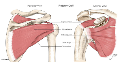
  • the rotator cuff is responsible for shoulder rotation, stabilization, and arm elevation
  • the rotator cuff holds the head of the humerus in the small shallow glenoid
  • rotator cuff muscles: supraspinatous, infraspinatous, teres minor, subscapularis
  • rotator cuff tear very common, 2 million people in US/year
  • important history in your diagnosis of rotator cuff injury: usually in dominant arm, age >40 years, pain worse at night, interferes with sleep, weakness with rotation and lifting, unable to do daily tasks (e.g. combing hair, putting on shirt)
  • Exam
  • acute traumatic usually occurs in setting of fall, trauma, significant amount of force (particularly in young patients)
  • chronic is degenerative, gradual onset, repetitive stress injury, occurs often in dominant arm, often as a result of bone spurs rubbing
    • >40% of people >65 have chronic rotator cuff tear
  • rotator cuff tears DO progress with time
    • full thickness tendon tears progress more rapidly
    • larger tears progress more quickly as well
  • Non-surgical management: activity modification, NSAID, cortisone?? (controversial, Dr. Pourtaheri doesn't recommend steroid injection for rotator cuff injury), PT helps with strength and pain, doesn't fix the tear, work on strengthening other tendons
  • All acute traumatic tears in people <60 should be fixed
  • "old tendons are not fixable" (no atrophy on MRI)
  • Shoulder arthroscopy: small incisions w/camera, nerve block for pain
  • Rotator cuff repair in correct candidates have 95% success rate (in terms of pain, function), improved shoulder strength and prevent tears from progressing
  • Post op course: 6 weeks in sling, 3 month limited lifting, PT

Many rotator cuff tears have an associated biceps tendon tear (should be repaired at the same time)

2) Impingement/Bursitis

  • inflammation of the bursa, usually due to overuse (overhead activity), sometimes trauma
  • pain exacerbated with activity, relieved with rest/NSAIDs, immobility
  • no-op treatment: activity modification, steroid injection, ice, ultrasound, PT
  • arthroscopic surgery for bursitis is generally arthroscopic bursectomy w/arthroplasty on the undersurface of the acromion 
  • rehab is faster than rotator cuff: sling x 2 weeks, PT within 2 weeks of surgery

3) Shoulder Labrum Tear

  • Labrum is fibro-cartilaginous ring that attaches to the glenoid, anchor point for gleno-humeral ligaments 
  • labrum is essential for shoulder stability in people <40
  • in people <40 tears are usually associated with trauma or dislocation event
  • in people >40, most labrum tears are physiologic and don't need treatment or surgery
  • PT is best non-operative management   
  • Sometimes surgery is indicated for people who are young and failed PT

4) Shoulder arthritis

  • >60 year old patient arthritis is a common cause of shoulder pain
    • articular cartilage thins out with time, exposed bone
  • X-ray: collapsed joint space, large bone spurs, thickening of subchondral
  • Non operative management: NSAID, PT, steroid/cortisone injection (yes, indicated)
    • PT to stretch the shoulder joint capsule (see exercises below)
  • Operative tx: shoulder arthroscopy (to release joint capsule, usually in mild to mod arthritis)) and shoulder replacement
  • Shoulder replacement 90% pain relief indicated for moderate-severe arthritis of gleno-humerus
  • there have been significant advances in shoulder replacement techniques and technology over the last 10 years
    • same day (outpatient surgery)
    • 4-6 weeks immobilized in sling, PT within a week, full recovery 6 months-1 year
  • two types of shoulder replacement: anatomic (intact rotator cuff) vs. reverse shoulder replacement (non anatomic)-- shoulder arthritis w/large rotator cuff tears
    • in reverse, ball goes on socket side of shoulder, socket on ball side of shoulder

Final pearls:

  • History and physical exam are key for assessment and diagnosis of shoulder pain
  • X-rays are still always a good idea as an initial evaluation (arthritis, calcific tendonitis, acromial bone spurs, for large rotator cuff tears for decision-making for surgery)
  • MRI definitively diagnose rotator cuff tears
  • Role for ultrasound? In patients who cannot get MRI (e.g. pacemaker), can use for shoulder injections (ultrasound guided)
  • Absolute indications for MRI in shoulder pain
    • fall/acute injury with sudden onset weakness in the arm likely has an acute rotator cuff tear(to evaluate for rotator cuff tear, which should be repaired within a couple months of injury for best outcome, time sensitivite)
    • if concern for biceps tendon "Popeye" arm (full thickness tear of biceps tendon)


Diverticular Disease (Sawyer, 8/11/2021)

Diverticulitis is. . . 

  • the 3rd most common cause of GI illness requiring hospitalization
  • the leading cause of elective colon surgery
  • a bit unpredictable but often managed medically
    • 15% of cases ultimately require surgery 
    • surgical indications: medical therapy failure (or not amenable)
Medical management
Medical/conservative treatment of uncomplicated diverticulitis generally involves antibiotics x 7-10 days
Note: there is NO high quality evidence regarding the ideal duration of antibiotics and which abx are superior
Good abx choices include: bactrim DS/flagyl (favorite of Dr. Sawyer), cipro/flagyl, levo/flagyl augmentin, and more

Diet
There is NO high quality evidence for specific dietary management of diverticulitis
Older surgeons prefer clear liquids x72 hours, advancing as tolerated after that (the rationale for this is really about possibility of surgery-- to be better prepared for a bowel prep if a surgery becomes necessary)
Of note, more recently trained surgeons will often let patients eat as tolerated (i.e. ad lib)
Of note, avoiding nuts, popcorn, seeds, corn, tomatoes, strawberries, etc is NOT evidence based and not necessary (JAMA 2008 paper). Do NOT tell patients to avoid these foods to prevent diverticulitis.

Known complications of diverticulitis:
  • Perforation
  • Fistula
  • Obstruction
Perforations that are very small are characterized as microperforations: conservative treatment with abx (oral/IV) is appropriate for microperforations; of note,19% of microperforations go on to form abscesses. Random pearl: diverticulitis as seen on CT often looks "worse" than the patient. 

Classification of perforation
  • Microperforation (not included in Hinchey classification system)
  • Hinchey I pericolic or mesenteric
  • Hinchey II walled off pelvic abscess
  • Hinchey III purulent ascites
  • Hinchey IV feculent ascites
Hinchey I and II can be managed non-operatively, III and IV should be managed with surgery

4cm abscess is used as the cutoff for IR drain placement
<4 cm diameter, abx alone usually sufficient 
>4cm diameter, IR drain + abx
if an abscess is exactly 4cm, it's at the discretion of the interventionalist/location of the abscess that determines best practice for management

What if it doesn't work?

If patients fail to improve after 48-72 hours of abx (usually repeat imaging is done), the next step is  a bowel preparation (if pt can tolerate it) and a surgical anastomosis. Of note, a "contaminated" peritoneum may require a diversion.

Surgical options:
colon resection w/primary anastomosis: one step, requires well-vascularized, non-edematous bowel, good nutritional status, good immune state (not immunocompromised)
colon resection w/proximal diversion
Hartman's procedure
open vs. minimally invasive: MIS preferred if feasible, shorter hospitalization/ileus and less pain. Long term, outcomes are about equal

Goal of surgical management of diverticulitis:
  1. Source control: remove the perforated segment
  2. Restore intestinal continuity-- depends on hemodynamics of the patient, degree of contamination, and surgeon preference/comfort
Free perforation (in an unstable patient) requires urgent damage control, which involves limited resection (if possible), peritoneal lavage, and usually a temporary abdominal closure. Alternative is a "Hartman's Procedure" with a limited resection, peritoneal lavage, end colostomy, and temporary closure.
  • in a study of 58 patients with generalized peritonitis and perforated diverticulitis, 9% mortality (5 patients). OF the 53 survivors, 44 were stoma free at 2 years (pretty good!)
Stable patient with feculent peritonitis (Hinchey IV) generally requires Hartman's procedure.
  • these can be difficult to close (only 50-60% have closure)
  • need to wait 6 months to 1 year for all the inflammation to resolve before rehooking


Of note, BOTH require a second look (return to the OR) at 24-48 hours

Fistulas can occur:
colo-vesicular 65%
colo-vaginal 25%
colo-enteric 7%
colo-uterine 3%

Obstruction: if diverticulitis is leading to obstruction, you MUST rule out cancer. Stenting-- while can be helpful in cancer-- is not helpful in diverticulitis

And, finally, the SSRRH Diverticulitis PROTOCOL
  • admit with IV abx
  • re-image at day 2-3
  • if improved on imaging--> abx management (7-14 days)
  • if imaging stable OR worse--> 
    • if drainable abscess, then IR drain, followed by surgery in 6-8 weeks,
    • if abscess NOT drainable, then mechanical bowel prep with surgery in 1-3 days

Vomiting in Children (Mueller, 4/21/2021)

Many thanks to Dr. Claudia Mueller, Stanford and CPMC pediatric surgeon, for an excellent presentation on Vomiting in Children-- her lens, unsurprisingly, was on the surgical causes of vomiting in children. 

As a family medicine physician, I don't typically consider vomiting in children a "surgical" problem, but it was sure a good reminder that sometimes it is! It's a hearty crew of clinicians who want to assemble at 7:30am to talk about vomit-- but hey-I have to tell you-- her presentation was excellent!  AND the best part was that Dr. Mueller gave us a number to call if we ever run into problems. 

To watch Dr. Mueller's excellent presentation click HERE.

For the Cliff's notes version, here you go:

  • Surgical causes of vomiting in children can rapidly progress to be life threatening
    • Ask yourself How sick is this kid? Do they have fever, tachycardia, moist music membranes, lethargy? Can I get them to stand for the KUB?
  • Presence of vomiting and ABSENCE of diarrhea is a concerning sign 
    • This makes sense; most vomiting in kids is related to acute viral gastroenteritis or food poisoning, both of which should be accompanied by diarrhea. The absence of diarrhea is a sign that surgical causes of vomiting should be on your ddx
  • The color of the vomit is key: color gives you some indication of the level the vomit is coming from (I know, I know, who wants to talk about the color of vomit) 
    • this is particularly true in infants
      • yellow/green (bilious) emesis in children <1 year is an "alarm bell that should be rung through the streets of any city" as it could be a surgical emergency (cardinal hallmark of a midgut volvulus that you do NOT want to miss)
    • most children will vomit food and other particulate matter, if they vomit long enough, they will eventually vomit bile, so prolonged vomiting leading to bilious vomiting may be less concerning than it starting out bilious
  • The intestine is a tube: in addition to the color of the vomit, what is coming out the bottom gives us a lot of information. If a child is having something out the bottom, they are much less likely to have true obstruction
    • Passing gas is best indication (more even than bowel movements)
SBO
  • Previous abdominal surgery is #1 cause of of adhesions causing SBO in children
    • traumatic surgeries (e.g. trauma ex-lap) are more likely to lead to adhesions
    • laparoscopic surgery maybe less risky (eg. laparoscopic appy) 
  • Farting is a good sign-- air doesn't just hang out in the colon; a child that is passing gas, even if there is an obstruction, it is at least partial
  • Be aware: not all kids with SBO get abdominal distention
  • An UPRIGHT KUB is the imaging modality of choice to evaluate for SBO in a child
    • want to be able to visualize: diaphragm, rectal gas
    • UPRIGHT is super important: air goes to top, liquid down to the bottom
      • air-fluid levels (straight lines) in SBO (can see in ileus, but more common in SBO)
      • a sick child who cannot stand up for KUB is concerning
    • CT scans can show more detail, e.g. the "point of the obstruction" but generally try to avoid CT scans in children <10 due to radiation
      • if you do CT scan, should do IV contrast; used to always require oral contrast (and can be more helpful), but should be done carefully due to risk of aspiration 

Upright KUB showing SBO
  • Initial treatment: NGT for decompression 
    • NGT should be adequate size (if it's too small, won't work as well). An NGT an actually treat SBO by relieving the pressure
      • Babies, size 10-12
      • Toddlers, size 12-14
      • age >7 years, size 14
      • teenagers/adults, minimum size 14, better >16
  • NGT has to be flushed, or it will get clogged
  • If NGT is working, as evidenced by the amount coming out of NGT decreases, and child starts feeling better, you may be able to avoid surgery
  • Another option after NGT: small bowel follow-through with gastrograffin (or ominpaque) can be diagnostic AND therapeutic
    • 25-50cc, repeat KUB 6-12 hours after administration: decreases hospital stay either because quicker to OR vs. able to discharge home
  • Hydration and serial abdominal exams are important in SBO
Midgut volvulus is the most urgent cause of bilious vomiting, usually in children <1 year old (85% before 6 months, 95% before 1 year)
  • A true emergency is caused because mesenteric vein and artery get twisted, no blood flow to the entire small bowel (colon and first/second part of duodenum have their own blood supply)
  • Can be life threatening in a few hours
  • Perfectly healthy baby totally fine, suddenly starts throwing up yellow/green, call a surgeon!
  • Imaging: UGI shows cutoff; x-ray may show just a stomach bubble (no other gas)
  • Consequence so dire: lose entire small intestine, may never be able to survive not on TPN

Pyloric Stenosis typically thickening/hypertrophy of pyloric muscle fibers
  • No one know why it happens
  • Usually age 2 weeks to 2 months, classically first-born males
  • Non-bilious (breastmilk or formula), progressive and persistent
  • Imaging: ultrasound
  • Surgery: cut open hypertrophic fibers, outer layer and spread it (pyloromyotomy)
  • Typically does not recur
Appendicitis
  • n/v, abdominal pain, umbilical down to RLQ
  • renewed interest in conservative management with antibiotics only
    • 95% of cases can be treated with antibiotics only, but 20% will recur in 1 year, 30% in 5 years
    • fecolith has VERY high recurrence, should be operated lap appendectomy
Ileocolic intussusception
  • generally age 6-36 months
  • small part of small intestine gets stuck in large intestine
  • usually due to laxity, lead point usually a lymph node, can be seen after enteritis OR after immunization (e.g. rotavirus vaccine)
  • Imaging: ultrasound, "target sign"
  • Reduction via radiology (air or contrast from anus into rectum, pushes the intussusception , reduces the small intestine), works large majority of time in kids without ischemia
    • 10% recurrence rate-->  to OR
  • Older kids need work up, lead point (e.g. lymphoma)
Hernias
  • bilious vomiting, if incarcerated
  • remember to take off vomiting baby's diaper to look for non-reduceable hernia

Abnormal Uterine Bleeding (Bartlett, 5/6/2020)

Thanks to Dr. Bartlett for her Grand Rounds this week on The Surgical Management of Abnormal Uterine Bleeding (AUB).

Here are some summary points:

Typical presentations of AUB:
  • irregular periods, heavy periods
  • abdominal cramping or dysmenorrhea
  • fatigue
  • dizziness
  • pelvic pain or pressure
  • missing work or school
AUB is subjective-- normal uterine bleeding is a 28 to 30 day cycle but definitions of "normal blood flow" is really based on a woman's "normal" menstrual cycle is.
Women should be treated for AUB if/when they request treatment and/or if it is adversely affecting their life--i.e. missing work or school. Unless a woman has symptomatic anemia due to her AUB, surgery is an elective procedure.

Diagnostic work-up AUB:
  • medical, surgical, and OB history
  • pelvic exam
  • pelvic ultrasound (BEST imaging for gyn organs)
  • CBC, TSH, pregnancy test (+coagulopathy workup particularly if young/teenage)
  • up to date pap smear
  • Endometrial biopsy advised (if >45 and/or risk factors)
Ddx:
PALM (structural)-COIEN (non-structural)
polyp, adnenomyosis, leiomyoma, malignancy or hyperplasia
coagulopathy, ovulatory dysfunction, endometrial, iatrogenic, not-yet-classified

Treatment options for AUB:
  • Combined oral contraceptives (birth control pills)
  • Tranexemic acid (TXA), used only during menses (slight increase risk of VTE)
  • Progesterone IUD (Mirena, skyla)
  • Hysteroscopy D&C
  • Uterine ablation
  • Myomectomy
  • Hysterectomy
Recovery from surgical intervention:
  • Hysteroscopy: outpatient surgery, bleeding and cramping, nothing vaginally x2 weeks
  • Uterine ablation: outpatient surgery, discolored vaginal discharge, nothing vaginally x 2 weeks
  • Myomectomy: depends on location (hysteroscopy vs. mini-laparotomy)
  • Hysterectomy: can be done outpatient (or overnight stay), most done laparascopically/robotically, pelvic organs removed through vagina IF cervix also removed


Small Bowel Obstruction (Sawyer 4/15/2020)

Big thanks to Dr. Russ Sawyer for an excellent presentation this week on Small Bowel Obstruction (SBO) during Grand Rounds. We are all getting better and better at this Zoom platform!

Cliff notes version of what primary care providers should know about SBO:
  1. Lactate is highly sensitive for SBO (but not that specific)
  2. The money is in the CT scan (CT is BOTH sensitive and specific)
    • with IV+oral contrast is are ideal, but IV contrast only is probably okay
  3. Many SBO patients don't need surgery (you actually can let the sun set on an SBO)
    • In fact, all patients NOT acutely ill (w/fever, leukocytosis, tachycardia) deserve a trial of non-operative management
    • SBO patients with a BM in last 24 hours likely will not need surgery
  4. Gastrograffin challenge is an effective way to differentiate patients who may need surgery vs. those who definitely don't. 
    • 90cc PO or via NGT x 8 hours in early/minimal symptom SBO (see below for details)
And now for the more robust version of my notes for those of you more in depth readers. . .

SBO is super common, >$1.5 billion per year in the US go to management of SBO

Classification of SBO
  • functional (i.e. adynamic ileus)
  • mechanical (acute vs. chronic, partial vs. complete) 
    • adhesions (account for 80% of SBO)
    • hernia (internal, groin, ventral)
    • malignancy
    • inflammatory disorders (IBD, ischemic bowel)
Pathophysiology of SBO
obstruction prevents people from passing food and air --> interluminal fermentation causes gas to accumulate--> bowel edema--> diminished absorption and decreased motility--> can gt transudative losses into the abdominal cavity (free fluid in the peritoneum)

Stats reminder from Dr. Sawyer, which is always helpful to review
Sensitivity: "positivity in disease" (how much you trust a positive result to mean the patient actually has that condition)
Specificity: "negativity in health" (how much you trust a negative result to mean that the patient actually does NOT have that condition).

(We are talking a fair bit about sensitivity and specificity this day with COVID testing. This is an excellent reminder!)

Clinical History
  • Acute abdominal pain (92%), usually precedes the onset of nausea/emesis
  • Nausea 
  • Emesis (82%-- more common than nausea)
  • Abdominal distention
Risk factors
  • Prior abdominal or pelvic surgery (even a simple appendicitis many years ago)
  • Abdominal wall or groin hernia (even internal hernia)
  • IBD
  • Prior irradiation of the abdomen
Physical exam
  • Dehydration (even if not apparent on labs), often notable on physical exam (dry mucous membrane, decreased skin turgor)
  • Abdominal distention (most important finding on exam)
  • Surgical scars
  • Tympanic abdomen
  • High pitched bowel sounds or more commonly a rush of bowel sounds
Labs
  • Leukocytosis is common
  • Electrolyte abnormalities (hyperNa, hypoK)
  • Lactate is helpful. It's extremely SENSITIVE 90-100%, Specificity 40-80%. This means that a positive lactate gives high likelihood of surgical SBO, but a normal lactate does not rule out an SBO
Imaging: "Everything in SBO comes down to imaging"
  • Plain film (KUB): not very specific or sensitive (equivocal 20-30% of the time, misleading in 10-20%), consider skipping it and going straight to the CT
  • CT with IV contrast (+oral contrast if possible-- oral contrast commits patient to 4 hours in the ER, often get get enough info with just IV contrast), 
    • >90% sensitive, 95% specific. So you can pretty much trust the CT. 
      • If the radiologist sees it, it's there. If the radiologist doesn't see it, it's not there.
    • CT can usually tell grade, severity and even etiology (adhesion vs. malignancy)
      • However, intra-operative location is only correct 60-70% of the time
  • MRI (if CT contraindicated, e.g. pregnancy)

Surgical vs. Non-surgical Management:
Patients with SBO should be evaluated for surgical intervention WHEN/IF they are acutely ill with fever, leukocytosis, tachycardia, metabolic acidosis, ongoing pain.

HOWEVER, without the above findings (or with only a few of them) MOST patients should undergo initial non-operative management (this includes both partial AND complete SBO). How so?

Management of early/minimal symptom SBO (with Gastrograffin)
For patients who meet these criteria, Dr. Sawyer and team are working on a protocol to be started soon

If patient meets the following criteria:
1) SBO on CT, 2) distention w/o emesis (x8 hours, or at least minimal emesis, only need NGT if emesis), 3) BM in the last 24 hours, 4) minimal leukocytosis (<14), lactate (<4), THEN you can give them a gastrograffin challenge

How do I do a gastrograffin challenge for minimal symptom SBO?
  1. Give patient 90cc of full strength gastrograffin (via NGT or PO);  order from pharmacy (not radiology)
  2. Wait 6-8 hours for BM (up to 24 hours). If they have plenty of BMs, they passed! You don't even need to do KUB
  3. If no stool in 8 hours, get KUB to look for gastrograffin. 
  4. If it has made it to the colon, can pull NGT, start clear liquids and probably let them go home. 
  5. If no contrast in cecum, repeat KUB next day. If in the colon, start clear liquids. If not, call surgery.
How does gastrograffin work? For this purpose, it is actually being used therapeutically (rather than diagnostically). Gastrograffin pulls fluid into the lumen and "flushes things through", decreasing the bowel wall edema and improving the SBO.

Who NOT to give gastrograffin challenge to? 
  • infection (e.g. appendicitis, diverticulitis)
  • cancer
  • incarcerated hernia
  • pregnancy
  • abdominal surgery in last 6 weeks
Prevention of SBO:
There is not great data on any intervention or product done intraoperatively to prevent adhesions and prevent SBO. However, if a patient has had a first episode of SBO or recurrent SBO, Dr. Sawyer recommends:
  • low fat diet (maintains intestinal transit time)
  • clear liquids are tolerated well (4-6 days): patients with SBO do not need to go home on a regular diet, once a patient feels true hunger, it's time to eat
Recurrence
  • 20% recurrence after first episode
  • after 3 episodes, their risk of recurrence is greater than 80%, need to consider surgery to lyse adhesions (depending on interval between the recurrences)


Diagnosis and Management of Osteoporosis (Hamann 7/23/2026)

 A recording of this presentation is available HERE .  *** Thanks so much to Dr. Kendal Hamann, SMGR Endocrinologist, for an outstanding Gra...