A recording of the presentation can be found HERE.

Showing posts with label plastic surgery. Show all posts
Showing posts with label plastic surgery. Show all posts
Skin Cancer Reconstruction (Pourtaheri 4/13/2022)
Many thanks to Dr. Navid Pourtaheri, new-to-our-community plastic surgeon, on Skin Cancer and Skin Cancer Reconstruction. This is a great summary of skin cancer findings and some specific indications and recommendations of when to involve plastic surgery.
A recording is available HERE
Skin cancer types: basal cell (BCC) squamous cell (SCC) keratoacanthoma, melanoma
Basal cell cancer (~4 million cases/year in US)

- variable in appearance, "can look like anything"-- don't know what it is until you remove it
- always slow growing (even high risk ones)
- primarily occur on sun-exposed areas (hand, ears, face)
- do sometimes spontaneous bleed or ulcerate
- can be locally invasive
- very rarely metastasize
Squamous cell cancer (SCC), 1.5 million cases/year in US

- variable appearance, but more commonly dry and scaly, also can be rough/thickened, wart-like, can ulcerate and form open sores
- more accelerated growth with local invasion than BCC (tend to progress more quickly)
- lower lips more common with SCC
- can be locally invasive, can metastasize
- curable if treated early
- actinic keratoses (AK) is pre-squamous cell lesion
Keratoacanthoma, 275K cases/year in US

- once considered subset of SCC
- dome-shaped lesion with central keratin plug
- almost always in sun exposed areas (face, ears, nose, hands)
- quite rapidly growing (faster than SCC), can be locally destructive
- unpredictable, can spontaneously regress
- less likely to metastasize
Melanoma, 197K cases/year in US
- most costly type of skin cancer
- 25% found in existing moles, 75% spontaneously occur on normal looking skin
- 50% are melanoma in situ>> high cure rate (90-100%)
- can be found ANYWHERE
- most common form is superficial spreading
- when goes deeper, called lentigo maligna
- 10-15% of cases are nodular melanoma, most aggressive, tends to be found invading
- remember ABCDE (see below) with special attention to "E" (evolving-- that is a changing mole)
- Breslow thickness correlates with 5-year survival


Treatment options for skin cancer
- Moh's surgery
- most commonly used in BCC and SCC (often contraindicated for melanoma)
- high risk areas (H zone)
- cosmetically sensitive areas, over joint surfaces
- decreases amount of tissue excised, maintaining the same cure rate
- Excision with margins and simple closure also acceptable
- Curettage (EDC)
- scrape the abnormal tissue, then burn it
- Cryotherapy more common in older patients, people who don't want a procedure, on blood thinners, large number (e.g. on face)
- Melanoma best excised w/margins, possible lymph node biopsy (plastic surgery, general surgery)
- sentinel node biopsy not indicated for melanoma in situ
When to refer to plastic surgeon?
- skin cancer not indicated for Mohs
- post dermatology resection, cannot close
- melanoma
Biopsy options
- shave: get full epidermis but only part of dermis (this is good because it won't form scar)
- always inappropriate for c/f melanoma because need full thickness
- punch: takes full thickness of skin
- incisional biopsy: get piece of abnormal with normal adjacent, done with scalpel
- excision: preferred for lesion is <1cm (cut the whole thing out with margin, 1mm of normal tissue is acceptable margin
- FNA not used for skin, but for clinically positive nodes
- Sentinel lymph node biopsy (SLNB) after melanoma diagnosis, after lymphoscintigraphy (radioactive dye that drains to the lymph node)
Imaging
- Always needed in >Stage 3 melanoma, definitely NOT indicated in stage 1 (stage 2, use your judgement, may be indicated)
- Most common: PET CT for assessing for metastases
- Chest CT also good modality b/c melanoma so often goes to lungs (chest xray not sufficient)
Wound Care (Cardenas, Cortez, Daly, 3/16/2022)
Many thanks to wound care nurse Wendy Cardenas, general surgeon Allen Cortez, and wound vac rep Kevin Daly for an informative talk this week on Wounds and Wound Care. I learned so much!
For a recording of their presentation click HERE (starting 11 minutes into the recording)
My notes:
- There are all kinds of wounds: surgical/traumatic, pressure, vascular, diabetic, infectious
- 1.3-3 million Americans are treated for pressure wounds each year
- 60,000 deaths per year are attributed to pressure wounds
- $9.1-11.6 billion dollars spent in the US on pressure wound are alone
- You only need 33 mmHg of pressure to create a pressure wound--> equivalent to a "gentle handshake"
Physician role in wound management
- decrease pressure
- decrease friction/shear forces
- manage incontinence (urinary, fecal>> indwelling/suprapubic catheters, ostomy)
- nutrition! nutrition! nutrition! (protein-calorie, blood sugar control, obesity)
- pain management
- managing comorbidities (DM, tobacco, drug addiction, venous stasis, etc)
- education (patient, caregiver, nursing staff, family)>> "you can tell people what to do, but if you give them the reason why, there is much more acceptance and compliance"
Initial management of wounds
- Identify and treat the cause
- Clean the wound--> tap water is just as good as sterile saline or any other wash (okay to shower day after surgery, no baths). Pulse evacuation (in OR) with fluid and pressure has been shown to improve outcomes. A simple syringe can also help w/debridement
- Keep wounds MOIST to allow capillary ingrowth (not too wet/not too dry)
- Debridement (wet to dry, enzymatic, surgical at bedside or in OR)
- you want to see bleeding
- Optimize nutrition (glucose control, protein)
- Optimize comorbidities (stop smoking!)
- Antibiotics if appropriate--> only if s/sx infection, "use common sense"
- Adjuncts (e.g. iodine/betadine-- can cause tissue damage, which can disrupt healthy granulation tissue. No evidence that adjuncts improve healing).
Q: What is the perfect wound dressing? A: Skin
Wound Vacs

What does a wound vac do?
reduces edema around wound
heals wound 60% faster than standard dressing
stretches cells--> leads to degranulation tissue
Contraindications to using a wound vac
No active cancer in the wound
Dead/necrotic tissue in the wound
Untreated osteomyelitis (if osteo is being treated, it is fine)
Dead/necrotic tissue in the wound
Untreated osteomyelitis (if osteo is being treated, it is fine)
Who qualifies for a wound vac at home?
if wound vac was started in the hospital, generally patients can go home with the vac, but a wound vac isn't always the best thing for a patient
wound vacs weigh 8-11 pounds, too heavy for some
need to have home health care (dressing change 3x/week, 1x/week has to be licensed person who measures the wound to demonstrate healing)
must be able to keep wound vac on 22 hours/day
- Of note, patients should be off their wound vac no more than 2 hours/day
- When vac dressings are used in skin grafts, you must use non-adherent dressing between the graft and the foam
- Vac instill: instills fluid (e.g. normal saline) into the wound. Protocol: dwell time 10 minutes, 2 hour suction. Vac instill leads to 40% more granulation tissue than regular wound vac. Only available in acute care (hospital, some LTAC/some SNF
- Prevena vac is used along closed clean incisions, helps approximate the wound. Not for everyone, only place in patients who are high risk for dehiscence (morbid obese, redo). Put on in the OR. Entire product is disposable. When stops holding suction, battery makes noise, it shoudl all get thrown away. No wound care needed, no home health required.
Prevena vac

Our Wound Care Queen, Wendy Cardenas, finished off the presentation with these gems
Four stages of wound healing: hemostasis (immediate)>> inflammatory stage (6 days, WBCs/macrophages debride bioburden) >>proliferation (up to 3 weeks, this is where wounds get stuck, go from acute to chronic)>>maturation (can last up to a year, skin is never going to be exactly the same, always a place that can reopen, tensile strength permanently compromised)
How to approach a wound ala Wendy Cardenas
1) Figure out what happened
chart review, look for all old notes pertaining to wound
ask the patient, "How did this happen?"
check for pressure points
2) Check for infection: is the surrounding tissue hot, red, indurated, painful?
wound bed funky, milky, shiny, smell bad, creamy or copious fluid
3) Foot wounds: pressure, diabetic, venous vs. arterial ulcers
venous: medial/lateral/posterior: irregularly shaped, shallow, yellow slough, not painful>> compress!
arterial: medial/lateral: round, deep, pale bed, don't bleed easily, not much pulse>> no pressure!
4) Wound products you might use in the hospital
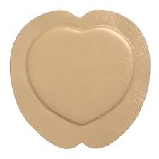
foam dressings: prophylactic on sacrum or coccyx, heel + offloading boot
plura-gel: adds hydration, cleans up wound
silver-impregnated hydro-gel
honey: better for superficial wounds
zinc: moisture associated breakdown, good for venous stasis legs (use w/compression)
Additional pearls:
- do NOT ever do superficial cultures on wounds; superficial cultures will only reveal polymicrobial organisms and skin flora
- quantitative tissue cultures are gold standard
- silver is bacteriostatic
- primary closure (clean wound), delayed primary closure (w/steristrips a day or two after), closure w/secondary intention
- skin graft wound vacs STINK when you first remove (5 days after a skin graft)
- if you have an abscess make a BIG BIG hole, making a tiny incision and packing will cause more pain. Big wounds are better
Big thank you to new-to-town-plastic surgeon, Dr. Melissa Mueller, for an excellent Grand Rounds presentation this week on Breast Reconstruction. I learned so much from her, and I look forward to working with her in our Sonoma County community.
A recording of Dr. Mueller's excellent presentation is available HERE. If you care for women in Sonoma County, you should definitely watch this.
Here are my notes:
- 1 in 8 women will develop breast cancer in their lifetime
- 80% of women w/breast cancer have breast conserving therapy (i.e. lumpectomy)
- of 20% of patients who undergo mastectomies, 40% get breast reconstruction, the majority implant based
- in SoCo, less than the national average of patients get reconstruction, likely due to lack of access to modern breast reconstruction in the area
- Breast reconstruction after cancer treatment is associated with improved self-image, decreased depression and anxiety, better body image, improved self esteem, and overall improved emotional, social and sexual functioning
- Some studies showing highest satisfaction with autologous reconstruction
- The 1998 Women's Rights and Cancer Rights Act guaranteed reconstruction for women getting mastectomy (national)
- California state law goes further and requires that ALL insurers (including Medi-Cal) must cover reconstructive surgeries (initial and subsequent) after breast cancer treatment, initial and subsequent implants (if patient needs an exchange)
- includes mastectomy AND lumpectomy
- includes contralateral breast symmetry procedures
- Breast reconstruction may be a "silver lining" to getting cancer

Type of breast reconstruction after breast cancer depends on type of surgery.
After Lumpectomy (i.e. Breast Conserving Therapy or Partial Mastectomy)
All women in CA are eligible for oncoplastic reconstruction as well as contralateral breast symmetry
To be eligible for a lumpectomy (vs. mastectomy), the size of breast cancer must be small in relation to the size of breast. After the lump is removed, there will be a hole in the breast, which needs to be filled. This can be done in two different ways:
- volume displacement: uses surrounding breast tissue to fill that hole--> that breast will be smaller and may require tissue rearrangement, reduction, lift/mastopexy
- volume replacement: brings adjacent tissue outside the breast to restore the original breast size (e.g. back, side wall/bra fat, implants)
- the contralateral breast will need to undergo symmetry procedure either immediately or delayed fashion
- this can happen at same time and/or after radiation
- radiation causes collateral damage and decreased breast volume by 10-15%, tightening of the skin, nipple uplift
- if reconstruction is happening before radiation, the breast that will be irradiated will be left slightly larger to allow for radiation changes
After Mastectomy, there are two categories of reconstruction
- implant based: most commonly performed with tissue expander placed at time of mastectomy. Expander is inflated q1-2 weeks until desired size is achieved, then is replaced with implant (saline, silicone)
- autologous: using patient's own tissue, make a breast mound with patient's own tissue, from abdomen/thigh/buttock/back (abdomen most common)

Reconstruction Timing
post-mastectomy reconstruction also can happen immediately (at time of mastectomy) vs. delayed
overall anesthesia time is less if doing delayed (in two parts)
if done delayed, another advantage is that final pathology is received (margins) to know if radiation will be required
of note, breast skin cannot be expanded after radiation treatment, so if a patient wants an implant-based reconstruction, they need to have fully inflated before radiation tx
in advanced cancer, pts should focus on adjuvent therapy prior to reconstruction
but for patients who have the choice, more immediate reconstruction is associated with better outcomes
Implant-based reconstruction
under vs. over pectoralis mm
historically, implant has been placed under the muscle because the tissue makes implant less visible
however, recently prepectoral breast reconstruction is gaining in popularity-- less pain, no animation deformity (doesn't move when pectoralis moves)
fat grafting (i.e. liposuction) can help make the implant less visible in both cases
for some women, surgeons can place implant at time of mastectomy--> fewer total surgeries
-have to have small breasts (A/B cup), want to stay same size, pts with nipple in correct place
-mastectomy is stressor to breast skin, so heavy implants can create a second stressor to the skin which can compromise blood flow to the skin
Autologous reconstruction:
create a breast mound using patient's own tissue (so you don't need an implant)
most common and most popular donor site is abdomen (deep inferior epigastric perforator flap)-- surgery similar to tummy tuck, kept alive by suturing to internal artery and vein in the chest
Fat grafting (ie liposuction--> injected with syringes after implant or autologous ( to disguise)

Counseling women for cosmetic results they may expect w/mastectomy
- chest wall can be concave after surgery
- lateral chest adiposity is not removed
- dog ears (standing cone deformity)
BIA-ALCL
A word on very rare condition/complication from breast implants called Breast Implant Associated Anaplastic Large Cell Lymphoma
associated between textured devices-- both textured tissue expanders and implants (these are no longer used)
Incidence of association: 1/30K implants placed--> 949 total cases worldwide
On average 8-10 years after implant (at least 1 year)
Sudden fluid collection (collecting within a matter of days) or a new mass associated with the capsule around the implant
There has been a recall on all textured implants
FDA doesn't currently recommend removing the devices because it is unknown if removing them decreases lymphoma risk, but if patients feel uncomfortable having the device in their body, insurance will cover their replacement or removal
Ultrasound vs. MRI to evaluate, aspirate 50cc of fluid should be sent for pathology and immunochemistry

Subscribe to:
Posts (Atom)
Diagnosis and Management of Osteoporosis (Hamann 7/23/2026)
A recording of this presentation is available HERE . *** Thanks so much to Dr. Kendal Hamann, SMGR Endocrinologist, for an outstanding Gra...

-
A recording of this presentation is available HERE . Many thanks to Dr. Andrew Brown, who gave an excellent Grand Rounds presentation this w...
-
 Great thanks to Dr. Jose Maldonado, a Stanford psychiatrist and neuropsychiatrist, who literally wrote the benzodiazepine-sparing alcohol wi...
