Showing posts with label cardiovascular disease. Show all posts

Cardio-Obstetrics (Soneji, 5/13/26)

A recording of this presentation is available HERE.

Thanks so much to Dr. Nisha Soneji, a new local expert in Cardio-Obstetrics! She joined SMGR in the fall and gave us a great presentation this week that was very family medicine friendly-- on the overlap of cardiovascular disease (hypertension, pre-E, valvular disease) and the peripartum time. It's a great presentation, and she is going to be an awesome resource to have here in our community.

"Pregnancy itself is a major stress test-- you are basically on a treadmill all the time".

US has a shocking rate of maternal mortality-- the highest among developed countries, 49.5/100K live births (highest for black women in the US) and CVD is the major contributor to maternal mortality. Significant racial and ethnic disparities are seen: black women 2.6x risk of death compared to white women. Advancing maternal age increases risk of maternal mortality (87.1 death/100K births)


CVD accounts for 33% of all maternal deaths. Whereas infectious risk of maternal mortality have decreased over the last decade, CVD related deaths are increasing, 2/3rds are considered preventable.

Most common cause of CVD death:
  • congenital heart disease
  • ischemic heart disease
  • valvular heart disease (esp stenotic disease: aortic stenosis, mitral stenosis)
  • hypertensive heart disease
  • congestive heart failure (peripartum cardiomyopathy, esp post partum)Contributing factors; delayed response to warnings (pregnancy symptoms mimic CVD), ineffective care, misdiagnosis, lack of continuity post partum (risk continues up to 6 months after delivery)
2/3 of maternal deaths occur during post partum period, when women tend to make less time to be seen (caring for newborn, going through hormonal changes, PP depression, etc)

CV changes during pregnancy: "Pregnancy itself is a major stress test-- you are basically on a treadmill all the time". If you are at risk for CVD, in can present in pregnancy or post partum.

See image below:
Normal findings in pregnancy: systolic murmur, elevated JVP, displaced apex, edema, increase in chambers on TTE, small pericardial effusion
NOT normal in pregnancy: S4, diastolic murmur, fixed splitting second heart sound, moderate to large pericardial effusionNOTABLY Unchanged in pregnancy: LVEF, REF, PASP


American College Cardiology


Who should be referred to Cardio-OB?
change in functional status
asthma not responsive to therapy
palpitations
chest pain/tightness that doesn't improve
syncope
SBP not controlled on med
oxygen saturation <90%
hx chemo can lead to HF in pregnant women (10% risk)
existing cardiac conditions: valvular disease, CHF


Risk Assessment:

Modified WHO 2.0 risk calculator


CARPREG

Who doesn't need referral: isolated sinus tachycardia, benign ectopy, mild hypertension managed on meds, normal BNP or TTE. If in doubt, refer!

Preconception counseling: high risk patients should get preconception counseling when risk of death is so high that they really should NOT get pregnant.
Assess risk
medication review (cannot use ACE/ARB)
genetic consultation (if CHD, increased risk of fetal CHD)Testing:
TTE: echo is first line monitoring tool
Troponin, BNP not routinely monitor, but good idea to check baseline if risk factors and/or symptoms. Can then compare post partum to antepartum BNP. Troponin can be ordered routine at the lab.BNP>200 can be normal in pregnancy
BNP >300 VERY suggestive of heart failure
CXR for shortness of breath
Treadmill stress tests in pregnancy can be done safely
Zio/holter
CT chest with angiogram can be considered if benefit>> risk
Hypertension in pregnancy
Chronic hypertension (<20 weeks), gestational hypertension (>20 weeks), preE (+organ dysfunction, Pre-Eclampsia with severe features, Eclampsia (with seizures)
BP Goal <140/90
Daily low dose ASA during pregnancy for preE/eclampsia prevention >12 weeks EGA in moderate to high risk patients
Benefit>>Risk
Pre E: 71% increased risk CVD, 2.5 risk CAD, 4x risk HF

Meds for BP: labetolol (shouldn't be used in asthma, decompensated cardiac function), can use nifedipine

Arrythmias
pregnancy increases aryrthmias due to increased blood flow and hormonal changes. Most common CV complication. Increases with age >41 years.
Preventable
SVT: vagal maneuvers, beta blockers, calcium channel blockers, digoxin (can be added if BB don't work) flecainide
Defer ablation to post partum (due to risk)
Afib: BB, digoxin, 2nd line calcium channel blockers, can be safely cardioverted
Can get implanted devices if need pacemaker

Heart Failure should be treated during pregnancy, cannot be deferred to post partum
-bad outcomes for moms and babies


Peripartum Cardiomyopathy
diagnosis of exclusion
new EF <45% without reversible cause
RF: maternal age, htn, preE, prior cardiomyopathy
present in 3rd trimester to 1 month (up to 6 months PP)
20% recurrence rate, contraception is important
risk of death 5-10% at 1 year
most people with recover EF, but future pregnancy brings higher risk
Meds: loop diuretic, hydralazine, isosorbide dinitrite, digoxin, beta blocker, ((IV dobutamine can be used), AVOID: ACE/ARB/ARNI, SGLT2
Vaginal delivery is recommended unless cardiogenic shock (safer than LTCS)

Valvular Disease
preconception counseling important, especially with L side valve disease (even if asymptomatic)
send to cardiology, need to get stress test pre-conception
TTE q trimester for mild-mod valve disease
R sided valvular disease (e.g. TR), need fetal echo, rarely need intervention>> vaginal delivery preferred
L sided valvular disease: regurgitation well-tolerated, stenotic disease NOT well tolerated in pregnancy (e.g. AS or MS, even if mild). At high risk for atrial arrhthmias

CAD in pregnancy
1/10K hospitalizations after pregnancy
Risk increases 3x
RF: age, black race, eclampsia/preE, known CAD, traditional risk factors (e.g. DM)
Spontaneous dissection (SCAD) most common cause of pregnancy-related MI (conservative tx recommended)

Perioperative Evaluation (Schneider - 10/4/23)

Sorry, there is no recording available for this session. 

***

Thank you to our very own Dr. Dave Schneider for an excellent presentation on Perioperative Evaluation. Unfortunately, I forgot to hit "record" on the zoom meeting, so we do not have a recorded version of his presentation. Sorry about that!

My notes:

First off, "clearance for surgery" is not our job!  Our job is to assess each patient's perioperative risks and optimize and manage those risk factors as they head into surgery. Do note that there are gender and race disparities in those who receive surgical interventions (BIPOC patients receive fewer PCI interventions, fewer orthoplasties and have increased mortality when undergoing these procedures).

The new-ish term for perioperative cardiovascular complications is Myocardial Injury after Non-Cardiac Surgery (aka MINS). 5-19% of surgical patients will experience MINS, 84% will be asymptomatic. MINS is associated with increased morbidity and mortality.

The American College of Cardiology (ACC) last updated their Guidelines for Perioperative Cardiovascular Evaluation and Management for patients undergoing non-cardiac surgery in 2014. These old guidelines are available here. The flow diagram is a doozy and the recommendations are confusing: 

In classic Schneider-fashion, Dr. Schneider invented an acronym to summarize their 2014 recommendations. It is called E-A-R-L-I

E: Emergent: if a surgery is emergent--> take patient to the OR and deal with the negative outcomes later

A: ACS: if the patient has s/sx of ACS, manage per guidelines

R: Risk assess --> use any of several tools (e.g. RCRI, NSQUIP calculator, MICA calculator, see below for more info)

L: Limitation of function --> if patient unable to  perform at 4 METs using the Duke Activity Scale, optimize their functional status before proceeding to surgery

I: Impact on decision? --> Yes or No? If the cardiac stress test outcome will change how/when you will proceed with surgery, then go ahead with a stress test. If not, then proceed to the OR.

The European Society of Cardiology (ESC) updated their guidelines on perioperative management more recently in 2022. And their guidelines are much more simple than those of the ACC. They are summarized here by the ACC. In essence, they say: 

Is the surgery. . .

Emergent? -->  Proceed to surgery without delay, cardiac testing is not feasible

Urgent? --> Proceed to surgery without unnecessary delay (using a multidisciplinary team to determine about individualized cardiac testing)

Time-Sensitive? --> Do the surgery ASAP

Their guidelines are summarized in this lovely flow charts. Don't you just love flow charts?


***

Should you check a Hemoglobin and Renal function in all patients pre-operatively? The answer is no, but your should check in intermediate and high risk patients. 

Don't forget, for everyone, advise smoking cessation!

There are two risks to consider in evaluating patients: 1) the risk of the surgery itself (e.g. highest risk includes intra-thoracic, vascular) and 2) the risk of the patient

  • If the surgery is low risk, no CV assessment needs to be made
  • If the surgery is intermediate risk, and the patient is either >65 or with CV risk factors, get an EKG and check functional capacity
  • If the surgery is HIGH risk, consider EKG and biomarkers** for patients older than 45, definitely get them for patients >65 or with CV risk factors. If the patient has known CVD, get a cardiology consultation and make a multidisciplinary decision. 
**Note: Biomarkers referred to above include BNP and/or Cardiac Troponin (also in table above). They have been shown to predict MI. Either one is predictive and do not change outcomes. 

There are several risk calculators to help you determine your patient's  risk level:
1) Revised Cardiac Risk Index (RCRI), which Dr. Schneider shortens to DRC4 (diabetes, risky surgery, CAD, CHF, CVD, Cr>2)
Each calculator is slightly different and variably useful depending on the patient in front of you. All three of these have been validated and you should get familiar with all of them.

Okay, now for a few pearls:
  • There is NO benefit to coronary revascularization before surgery.
  • Labs and other tests should only be done pre-operatively if you were going to do them anyway
  • Coag testing is usually unnecessary unless patient is on warfarin. Family Hx and PMH are just as predictive of bleeding (e.g. if patient has history of prior bleed, or family member has bleeding problem, patient has higher risk of bleed)
  • Only get a pre-op EKG if the patient has known CVD, CV risk factors >65 and they are having an intermediate/high risk surgery
  • TTE only needed if patient has known valvular lesion and no TTE in the last year. You may consider if new onset dyspnea or change in status of their HF
  • Pre-op CXR is NOT recommended (Choosing Wisely, ACR 2017)
What about medications?
Statins: if a patient is on a statin, continue it (okay to miss a few days due to NPO, etc.). Perioperative initiation is reasonable if someone is getting vascular surgery

Beta blockers: if patient is already on BB, continue them perioperatively (perioperative withdrawal has 4x increased mortality). You may consider decreasing BB dose due to risk of hypotension after surgery. You can consider starting a BB at least one week (up to 28 days) prior to cardiac surgery if high risk patient and high risk surgery. 

Other anti-hypertensives: post-op hypotension is a common problem. Consider holding all BP meds on day of surgery, add them back slowly post-op, ?one at a time

ASA: it is okay to go to the OR on aspirin. Also okay to stop ASA in high bleeding risk patients (e.g. those on DOAC or warfarin as well). Continuing ASA has been shown to be cardioprotective: decreased MI by 56% and decreased composite CV outcomes. There is a non-significant increased bleeding risk if you continue ASA.

What about patients with recent drug eluting stents (DES)? Delay elective surgery for up to 6 months if possible so as not to interrupt DAPT. 


Thriving or Surviving: The Connection Between Chronic Stress, Chronic Disease, and Social Determinants of Health (Deol - 9/6/23)

 A recording of this presentation can be viewed HERE

***

Thank you to Dr. Navi Deol, PGY3, who gave an excellent presentation this week titled Thriving or Surviving: the intersection between chronic stress, chronic disease, and the social determinants of health. Each of the items in the subtitle is a HUGE topic, and Dr. Deol was able to weave them together beautifully and powerfully. I encourage you to watch yourself.

Image from https://www.glasbergen.com/stress-management-cartoons/cartoons/page/3

If you prefer the written word, see my notes:

  • stress: how certain stimuli (stressors) affect a person's mind, body and spirit
  • stress response: how our body reacts normal, a normal physiologic and psychologic response to stressors
  • some stressors evoke positive emotions and can be beneficial (eustress); some evoke negative emotions and cause problems (distress)
A certain amount of stress is important and necessary to generate optimum productivity and performance, but too much stress can lead to anxiety, overload, and burnout (see image below of the Yerke's-Dodson Law).
Yerke's-Dodson Law, image fromhttps://stock.adobe.com/

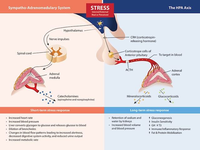
Stress causes physiologic changes in our bodies. We are all familiar with the autonomic nervous system, which responds to acute stress with the sympathetic "fight or flight" and the balancing parasympathetic "rest and digest", but what happens when the stress response is constantly being activated? 

Image from: https://www.backtothebooknutrition.com/adrenal-fatigue-hpa-axis-dysregulation/


The answer is that our long-term stress response leads to a cascade of responses that make us more vulnerable to chronic diseases.

A 2019 study published in the Journal of ACC looked at the link between SES factors such as low income and higher crime on MACEs (cardiac death, myocardial infarction, unstable angina, cerebrovascular accident, peripheral artery disease with revascularization, or heart failure). This study suggests that a biological pathway contributes to this link, involving, in series, higher amygdala activation, increased activation of the bone marrow (with release of inflammatory cells), which in turn leads to increased atherosclerotic inflammation and its atherothrombotic manifestations


Patients were categorized according to quartiles of their neighborhood median income and neighborhood crime rates.  Amygdalar activity (A) and arterial inflammation (B) were lower as neighborhood median income increased. Amygdalar activity was higher (C) and arterial inflammation trended toward an increase (D) as neighborhood crime rate increased. One image from that study is seen below. For more information, click the link above or see the study link below the image.

https://www.jacc.org/doi/10.1016/j.jacc.2019.04.042


Chronic diseases are non-communicable illnesses that persist for long periods of time and result from a combination of genetic, environmental, and psychological factors. These include cardiovascular disease (e.g. hypertension, coronary artery disease, and strokes), metabolic disorders (e.g. type 2 diabetes and obesity), mental health issues (e.g. depression, generalized anxiety disorder), and substance use disorders.

We know that inflammatory cascades play an important (and damaging role) in the onset and progression of chronic disease. While acute inflammation is technically "good" for us because it cleans up disease states in our body, chronic inflammation is bad bad bad.

Financial stress

Are you aware that money (finances and inflation) is a tremendous source of chronic stress for a shocking number of US adults. A 2022 survey found that 57% of US adults reported not having enough money to pay for essential items; 43% reported that they are not saving enough, and 56% had to make different choices due to their lack of money. 

Violent stress

Mass shootings, gun violence, and crime are also sources of tremendous chronic stress, particularly for people living in poverty and disproportionately for BIPOC people. 

Social Determinants

This leads us directly into a discussion of the importance of the Social determinants of health (SDOH), the conditions into which individuals are born, grow, live, work and age. There is an unsurprising link between chronic stress and the SDOH. These include your neighborhood and built environment, healthcare, education, economic stability, and social and community context. 

image from Healthy People 2030

If this is news to you, check out this video about how zip codes influences an individual's health: A Tale of Two Zip codes.

And for something even closer to home, check our our local Sonoma County data on how the SDOH vary based on zip code in the report titled A Portrait of Sonoma County 2021 Update, available here:  https://upstreaminvestments.org/impact-make-a-change/portrait-of-sonoma-county

https://upstreaminvestments.org/impact-make-a-change/portrait-of-sonoma-county


What can we do about all this stress?
Dr. Deol encouraged us to take a deep breath and realize that we cannot tackle these complex issues alone. First, we must acknowledge the deep-rooted history of structural and systemic racism, oppression, and discrimination that have led to health inequities that require interventions at multiple levels to reduce disparities. 

When caring for individuals, we should be careful about using the term "non-compliance" and better recognize the daily barriers our patients face due to their own SDOH. In our communities, we should be screening for SDOH and get to know and refer to appropriate community services. And at the state level, she encouraged us to support CAFP Bill AB85, which requires SDOH screening and provides resources and education for providers in referring to community health workers. 

More information for AB85 can be found HERE


Understanding Methamphetamine Use Disorder (Freschl 8/9/23)

Many, many thanks to Dr. Guille Freschl, who gave Grand Rounds this week titled Understanding Methamphetamine Use Disorder: A Deep Dive.  This was our first R3 Grand Rounds Presentation of the academic year, and Dr. Freschl knocked it out of the park. The link to a video recording of her presentation is available here. Below find my notes.   

A recording of this presentation can be viewed HERE.

***

Dr. Freschl was motivated to present on this topic by a longstanding interest in substance use disorders coupled with curiosity and concern about the oft uttered "Oh, it's probably because of the meth" that she heard from the mouths of her teachers. She was left wondering where the science meets the bias.

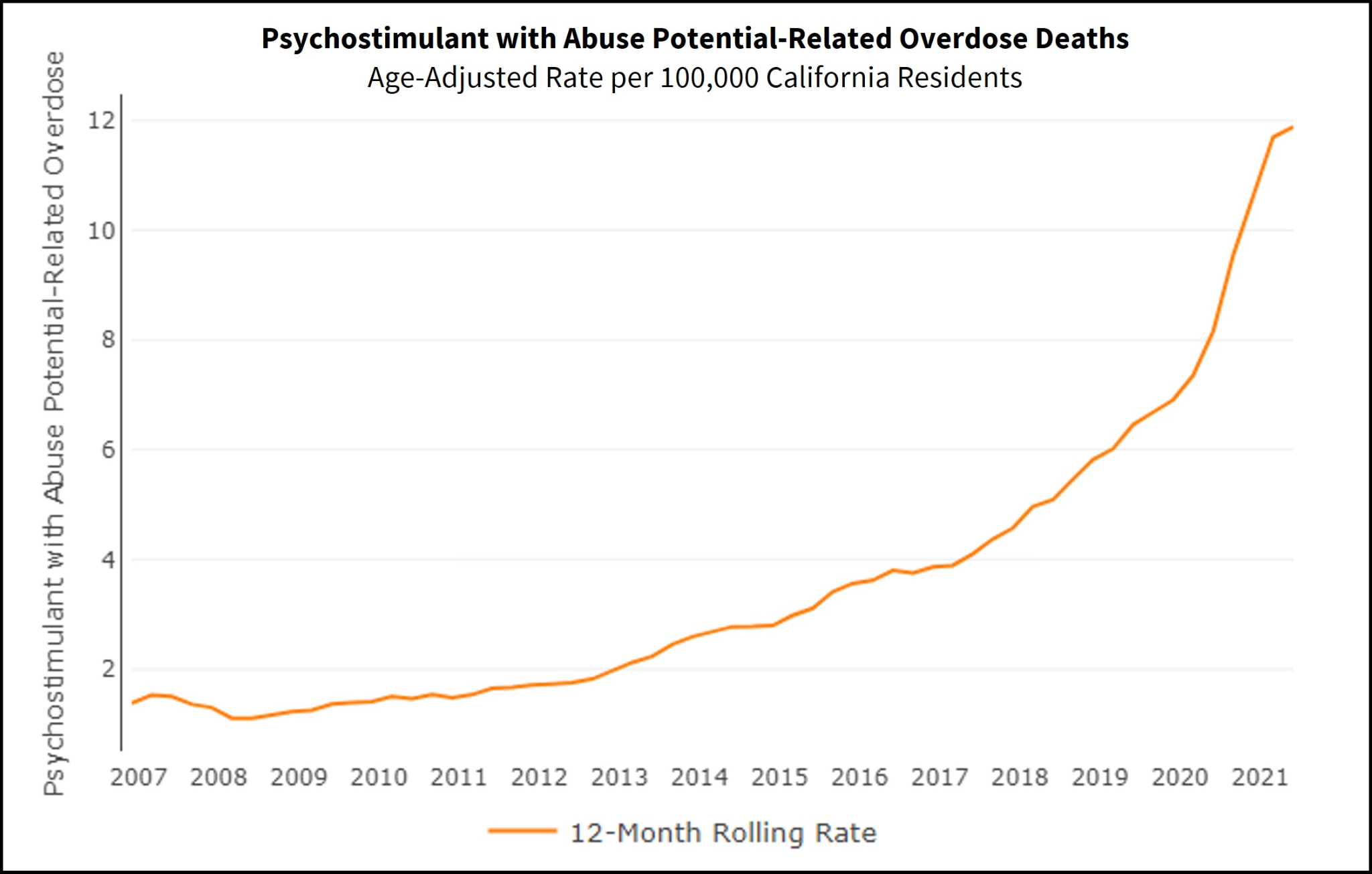
Did you know that amphetamine-type stimulants are the most widely used drugs in the world after cannabis?  Did you know that  between 2011 and 2016, overdoses from methamphetamine TRIPLED and that 1/4 of all overdoses in 2021 in the US were due to meth?

Methamphetamine use disorder can be seen all over the nation, but prevalence varies per region. Rates are highest in the West Coast and South. For example, prevalence of reported meth use in the past year in CA is reported at 1.04% of all adults, almost twice as much as most states in  the Northeast (see map below).
 
In California, non-fatal ED visits and overdose deaths have both risen over the last decade. In fact 32% of those in court-mandated substance use disorder treatment programs were there due to methamphetamine use. While the bulk of media and political attention is currently focused on opiates, one wonders, why aren't we talking more publically about methamphetamine?


What is methamphetamine?
Methamphetamine is an amphetamine derivative, notable for its additional methyl group; it enhances dopamine and norepinephrine in the synaptic cleft. Meth has a very long half life (12 hours cmpared to 90 minutes for cocaine). 

Why is meth bad? So many reasons. . . keep reading to understand a few of the major adverse effects. 

Cardiovascular toxicity

CV toxicity is the #1 cause of death in patients using methamphetamines, and risk of sudden cardiac death is increased by 27% with active meth use. CV toxicity includes a range of end-organ issues, including:
1) Hemorrhagic and ischemic strokes, due to vasoconstrictive effects and cerebral hypoperfusion
2) Very high rates of coronary artery disease (CAD) -- half of patients with regular meth use have CAD, despite lower rates of obesity and diabetes in these patients. This is thought to be directly related to the pro-inflammatory effects of meth. 
3) Angina, which does not respond well to nitroglycerin, is common, due to vasospasm
4) Pulmonary hypertension, especially with IV meth use, due to damage to pulmonary endothelial cells
5) Severe systolic dysfunction with LV dysfunction is another sequalae of meth use
6) Ventricular arrhythmias are notable

Neurotoxicity
Neurotoxicity is the #2 cause of morbidity and mortality in patients using meth. It rapidly crosses the blood brain barrier. It does a doozy on the brain, including disrupting pleasure centers, creating episodic memory issues, damaging executive function (2/3 of people with regular meth use show cognitive impairment, worse with older age and longer duration and frequency of use), disrupting motor function (including fine motor and choreas), and can lead to psychosis similar to schizophrenia (delusions of persecution, auditory hallucinations, and formication in almost half of people using). 

There is also a direct relationship between meth use and Parkinson's disease.

Dental effects
Serious dental effects include caries, tooth loss, tooth fractures -- all due to decreased saliva production (xerostomia), teeth grinding and jaw clenching that occurs with meth use.

Medication Assisted Therapy (MAT)?
Unfortunately, there are no FDA approved treatments for methamphetamine use disorder. A large meta-analysis of 43 RCTs with over 4000 patients found no clear evidence-based effective treatment. 

These included trials with mirtazapine (conflicting results), methylphenidate, bupropion, naltrexone and modafinil (limited evidence of benefit, no support for routine use). In addition, anticonvulsants, antidepressants, antipsychotics all low strength and insufficient evidence. Bummer. 

There was a small study that suggests that methylphenidate may be associated with decreased use over time: no difference at 30 days, but decreased in self reported use days at 10 weeks. 

Also, a small study of combination therapy --  IM naltrexone (380mg q3 weeks) PLUS PO bupropion (450mg daily) small treatment effect of 11% reduction in meth use. 

Hopefully, people will continue to investigate different agents for MAT and treatment of meth use disorder!

In conclusion, Dr. Freschl recommended that we use shared decision-making with patients when talking about trialing non-FDA approved treatment options. She reminded us to screen for CV and neurological sequelae of methamphetamine use. 

Many thanks to dynamic duo podiatry team, Drs. Walter D'Costa and Kevin Grierson, for their collaborative care of patients and for their Grand Rounds presentation on Everything Foot this week.

A recording of their presentation is available HERE .

Dr. D'Costa started the presentation with the practice changing pearl of the day: 

***Remind our patients (at high risk for foot problems) to change their shoes 1-2 times per day to relieve pressure.***


This is such a great pearl-- definitely not part of my regular prevention spiel, but an easy daily practice that can help prevent chronic friction issues with the same pair of shoes.

A review of the three common categories of foot ulcers: neuropathic, vascular, and ischemic.

1) Neuropathic ulcers result from insensitivity (i.e. neuropathy), inability to perceive pain, which leads patients to walk on bony prominences, and then to get skin breakdown, which can become chronic. Neuropathic ulcers present with some key features:

  • hypertrophic rim of callous
  • fibrotic wound bed
  • painless
  • not very much necrosis (compared to vascular ulcers)

  • The heel is a common site of pressure (in patients with heel ulcers, don't forget to screen for restless leg syndrome, which can lead patients to rub heels and lead to neuropathic ulcers)
Prevention:
  • diabetic foot exams
  • patient doing daily foot check
  • changing shoes frequently
Treatment:
  • OFFLOADING is key (inserts, change in shoe, etc)
  • Aggressive debridement by podiatry, wound vac as needed
  • Good diabetes control
  • Sometimes excision of the bony prominence
2) Venous Stasis Ulcers result from incompetent valves and mast cell inflammation, leading to skin breakdown, they often occur at medial/lateral malleoli
  • brown discoloration (stasis), chronic edema (often decades)
  • dry skin--> scratch--> opening/fissures--> infection--> infected ulcers (pearl: Make sure patients with venous stasis hydrate their skin daily with lotion/cream, even baby oil)
  • usually these do not have hyperkeratotic margins
  • these often weep (and weep and weep)


Prevention:
  • Control EDEMA via compression stockings, diuretics, elevation, venous pump/sequential pump (these pumps are DME covered by most insurances, particularly if patient has chronic stasis and/or hx of an ulcer)
  • lotions to keep skin moist
Treatment:
  • Sharp debridement by podiatry
  • Enzymatic dressings, wet-to-dry dressing, calcium alginate (absorptive of weeping), hydrocolloid, silver-impregnated gauze
  • Antibiotics if infected
  • Biopsy the ulcer (if don't improve with good treatment)
  • Grafting
3) Ischemic Ulcers are almost always very PAINFUL (unlike neuropathic and venous stasis), dark necrotic tissue


Treatment
  • NEED revascularization
Additional foot ulcer pearls:
  • For heel decubitus ulcers, always get x-ray to rule out osteomyelitis because these are by definition unstageable 
  • If you see a red hot foot in a diabetic, don't forget charcot arthropathy: red/hot/swollen foot, "rocker bottom" must be treated with immobilization, can appear like acute infection (elevated WBC, ESR, but these don't improve with abx)
  • Edema Wear has a number of excellent products, including open toe stockinettes, for compression products that may be more useful to patients who have trouble using compression stockings.
  • Also consider less rather than more compression if the patient is not going to wear compression stockings at all. Some is better than none. 
For part 2 of the presentation, Dr. Grierson covered several key toenail diagnoses including ingrown toenails, pigmented toenail lesions, onychomycosis, and subungual hematomas

1) Ingrown toenails are super common-- 20% of primary care foot complaints. Usually occur in younger patients, a result of trauma, improper cutting, tight shoes, and hypertrophic nail folds

Lifestyle advice: avoid tight shoes, warm water soaks for early symptoms 
For mild ingrown nails: oral antibiotics, gutter splints

Surgical treatment includes: partial vs total nail avulsion with or without chemical matrixectomy. Of note, partial nail avulsion has a 39% recurrence rate and total nail avulsion has a 83% recurrence. HOWEVER, Practice changing pearl:

*** Chemical matrixectomy with toenail avulsion (e.g. 88% phenol) has a 3% recurrence rate. You definitely should be doing a matrixectomy if you are removing a toenail***

Many patients complain about their toenail removals: they were so painful, miserable, inadequate anesthesia. A word on nerve blocks: the most important nerves to numb up the toe are on the plantar surface. For good anesthesia, Dr. Grierson recommends a ring block with ~3ml of lidocaine (1 or 2% w/o epinephrine). This should be injected into the SUBCUTANEOUS space and if you're in the right space, there should be very little resistance, i.e it should go in easily, causing the patient little distress.

2) Pigmented lesions in nails (longitudinal melanonychia) are common and have a long ddx: this includes ethnic variation, pregnancy, drugs, chronic local trauma, endocrine abnormalities, and the big bad wolf: melanoma

Ethnic melanonychia is very common in people of color of all ages, but increasing incidence with age. In fact, studies show a 20% incidence in people of Japanese descent and up to 100% incidence in African Americans over age 50.

Warning signs for melanoma of the toenail:
  • single nail (usually the large toe)
  • >3mm width of pigmented band
  • more irregular border
  • recent changes (e.g. increase in size, rapid growth)
  • family history of melanoma
  • Hutchinson's sign: pigment in the nail extends to the nail fold
  • benign ethnic melanonychia

    subungual melanoma
If in doubt, refer for biopsy (btw pigmented lesion biopsy needs to come from the nail matrix)

3) Onychomycosis is a common dermatophyte infection of the toenail, affecting 10% of the general population, 20% of people >60 and 50% of people >70. It causes discoloration, thickening of the toenail and can lead to other chronic foot problems

Treatment:
  • Debridement (symptomatic relief)
  • Topical medications: tavaborole 5%, ciclopirox 8% lacquer don't have high efficacy rates but can work for some patients
  • Oral medication: terbinafine (Lamisil), itraconazole
    • mycologic cure rate for terbinafine is 70%, itraconazole 54%
    • complete cure 38% for terbinafine, 14% for itraconazole
    • elevation in AST/ALT is VERY rare with terbinafine, <1% and generally self resolve, serious life transaminitis is even more rare 1/500K-1/120K
  • Alternative therapies for onychomycosis include apple cider vinegar, tea tree oil.There aren't great studies, but apple cider vinegar does contain maleic acid, which has fungicidal properties, and tea tree oil may have synergistic effect with topical antifungals
4) Subungual hematomas occur as a result of trauma. 
  • Fracture is common (10-25% of people w/associated phalanx fracture). For this reason, these toes should get x-rayed. 
  • For symptom relief, trephination (cool word, definition: to open with a hole saw (i.e. trephine)) with a simple 18 g needle is safe and effective (spin, painless, no anesthesia required). You may need to make more than one hole. Go for it!


Pulmonary Hypertension (Wang, 1/13/2021)

Dr. Helena Wang gave an excellent Grand Rounds this week on Pulmonary Hypertension. 

You know when you have read about a particular topic a hundred times and you still don't quite grasp even the most basic of concepts? Well, that is pretty much how I have felt about pulmonary hypertension for the last decade. I get all tangled up in the RVSP and the strange WHO Classifications and have never quite been able to solidify my illness script for pulmonary hypertension. 

Well, Dr. Wang, thank you. I feel like I am finally here. Pulmonary hypertension is such an interesting intersection between the lungs and the heart and a good opportunity to review basic physiology too! As one of our residents said to me yesterday after the talk, " I feel like hers is a presentation I want to watch again and again." Me too (and I did!). For those of you who want the written summary, here goes:

First a refresher on Pulmonary Function Tests (PFTs):

  • The FEV1/FVC Ratio should be used to determine if a patient has an OBSTRUCTIVE lung disease. 
    • Remember that an FEV1/FVC <70% is consistent with an obstructive disorder (e.g. asthma, COPD, etc)
  • Total Lung Capacity (TLC) is used to decide if a patient has a RESTRICTIVE lung disease
    • 80-120% is considered normal 
    • Patients with restrictive lung disease will have a TLC <80%
  • Diffusion capacity for carbon monoxide (DLCO) provides information on the efficiency of gas transfer from alveolar air into the bloodstream
    • for these defects in gas exchange, ddx includes pulmonary hypertension and liver disease

Second, reminds the pulmonologist, don't forget to pay attention to the right side of the heart in the echocardiogram!!  Dr. Wang urged us to scroll down and look at the text. Is the RV big? What is the velocity of the regurgitant jet? What is the TAPSE?


  • A first hint of pulmonary hypertension on a TTE comes on apical 4 chamber view where the R ventricle shows turbulent flow back into the R atrium
  • RVSP (right ventricular systolic pressure) is an estimate reported on most echo reports and can be very challenging for the Echo tech to calculate
    • First they measure the velocity of tricuspid regurgitant jet (velocity >3 is a clue that there may be pulmonary hypertension present)
    • then they guestimate of CVP (0, 5, 10) based on compressibility of IVC
    • NOTE: a calculated RVSP>25 is considered HIGH right sided pressure
  • The R ventricle is often very plump and dilated under high pressure (so. . .a big chubby RV on echo is another clue that you might have pulmonary hypertension)
  • Tricuspid Annular Plane Systolic Excursion (TAPSE): RV contracts in a twisting motion with each contraction as RV shortens. Normal excursion is 1.5cm. 
    • If RV is volume overloaded and dilated, cannot squeeze as well, not shortening, TAPSE drops <1.5cm on echo
And finally, right heart catheterization is gold standard for measuring R sided pressures, but really this should be done after you have done a full evaluation (see below) once you are pretty certain you have primary pulmonary hypertension (WHO Group 1)


WHO Classification of Pulmonary Hypertension
  • WHO Class 1: Pulmonary ARTERIAL Hypertension (it's like going from a 6 lane highway down to a 2 lane highway--> pressure goes up, fluid backs up)
    • WHO Class 1 has the worst outcome: 5 year survival 61%
    • only once other causes of pulmonary hypertension are identified and treated
    • Seen in HIV (regardless of viral load, duration), Connective tissue disease (e.g. scleroderma), portal hypertension
  • All other WHO classes are actually pulmonary VENOUS hypertension (something downstream to the lungs causing poor forward flow, back up of blood, then high pressure on R side of heart). These include
    • WHO 2: L heart disease
    • WHO 3: bad lung disease or hypoxia
    • WHO 4: chronic thromboembolic pulmonary hypertension (small, little tiny chronic)
    • WHO 5: potpourri
Once Pulmonary Hypertension is identified, the next question is DOES the patient have pulmonary hypertension that is primary or secondary

Ddx pulmonary arterial hypertension (WHO Group 1)
  • idiopathic
  • heritable (no current gene therapies)
  • drug and toxin induced (fen fen, methamphetamines including stimulant medications prescribed)
  • connective tissue disease (scleroderma)
  • HIV
  • portal hypertension
  • congenital heart disease
  • Schistosomiasis (#1 cause of pulmonary hypertension outside the US)
Ddx WHO Group 2: a bad heart
  • left heart disease: LV dysfunction (systolic and diastolic), valvular dz, outflow obstruction
Ddx WHO group 3: bad lungs
  • asthma and COPD (need PFT: spirometry, lung volumes, diffusion capacity)
  • ILD (need HIGH resolution CT chest)
  • sleep disordered breathing (episodic hypoxia at night causes vasospasm), need sleep study
  • chronic high altitude
  • developmental lung dz

A quick interruption to review the three types of CT chest you might consider to evaluate the lungs:
  • CT angiogram (aka CTA) is a very quick scan from feet to head chasing the dye, itskips very low bases and apices of lungs and should never be used to assess for interstitial lung disease
  • A High Resolution CT Chest: much thinner cuts (2mm) than a CTA starting at apices and going all the way to the bases. 
    • Very high detail of parenchyma
    • Ground glass opacities can be atelectasis vs. ILD--> flip when prone to see if atelectasis resolves while doing high res scan
  • CT w/wo contrast (e.g. nodule) 
    • can see 4 generations of branches of bronchial tree, but keep in mind that there are 17, so you cannot see filling defects in the small peripheral vessels

    Ddx WHO group 4: chronic thromboembolic pulmonary hypertension (itty bitty clots on VQ scan)
    • need VQ scan to get the level of detail in microvasculature
    Ddx WHO group 5: potpourri
    • hematological disorder (myeloproliferative)
    • systemic disorders (sarcoid, LAM--> chylothorax)
    • glycogen storage disorder
    • thyroid disorders
    • chronic renal failure
    • mechanical (fibrosing mediastinitis, tumoral obstruction)

    Right heart catheterization

    Once you have maximally treated all WHO 2-5 diagnoses, treat as aggressively as possible (e.g. diastolic dysfunction, COPD/asthma, chronic clots, sleep apnea, etc), then you repeat ECHO and see if there is still elevated pressure on R side of heart--> NOW, patient should get a right heart cath to confirm exactly what the pressure is.
    • On A R heart cath, PA pressure normal <25/15
      • Mean PA pressure normal is <25
      • Pulmonary hypertension = mean pressure >25
    • Gold standard is also to do a vasodilator challenge during R heart cath to see if there is reversible spasm in the pulmonary arteries? (inhaled nitrous oxide, epoprostenol, or adenosine--> look for decreased pressure)
      • if PA pressure decreases by 10 mmg HG, ending pressure <40, no drop in cardiac output
      • less than 10% of patients have + response to vasodilator
      • calcium channel blockers are treatment of choice for those who do have vasospasm
    When to treat Pulmonary Arterial Hypertension?
    There is NO number for pressure goals. Look at patients functional class, 6 minute walk, and Echo
    Treatment goals:
    • Functional class: symptoms at rest (class IV), normal (class I): goal is 2 or less
    • 6 minute walk test: goal is 400mg in 6 minutes ("please walk as far as you can in 6 minutes")
    • BNP: can be elevated in R heart strain, goal is normal
    • Echo: goal is RV not fat and chubby, decompressed and slender, TAPSE moving nicely
    Pulmonary Hypertension Therapy

    • Endothelin pathway: block (bosentan (black box liver toxicity, no longer used), ambrisentan, macicentan: side effects: edema, check LFTs, watch for rare malignant facial edema
    • Nitric oxide pathway: enhance (sildenafil TID, tadalafil QD, no lab monitoring, can get blue green colorblindness, rare complication nonarterici anterior ischemic optic neuropathy (sudden blindness one eye, reversible when stop), riociguat (TID, more systemic hypotension)
    • Prostacyclin pathway: enhance (potent, short half life and unstable)
      • epoprostenol: IV infusion, tubing connected to a pump, best mortality data, very labor intensive
      • treprostinil: IV vs. sq (like insulin pump) and inhaled QID (takes a lot of time, breathing treatment like nebulizer, 20-30 minutes per treatment
      • iloprost: inhaled 6-9 times/day
      • selexipag: oral BID, no labs but unrealistic uptitration protocol
    Guidelines for treatment are based on functional class. The ultimate goal is minimal symptoms with regular activity
    If functional class IV-->  needs prostacyclins right away
    If functional class III-->  can start with orals (e.g. tadalafil and macicentan)

    Any questions? Email or staff message Dr. Helena Wang at: wanghl@sutterhealth.org


    Cardiovascular Benefit of New Diabetes Medications (Magnotti, 4/22/2020)

    One of the reasons I love Grand Rounds is that each week I know-- whatever the topic, whomever the speaker-- I will walk away wiser. My curiosity will have been piqued. I will have learned something. I will have been challenged.  I will be a better family physician. And I will have often have new (more educated) questions.

    And as expected, this week, Dr. Mike Magnotti (SMGR Endocrinology) did not disappoint. He gave an excellent presentation on the cardiovascular benefits of two classes of "new" diabetic medications: GLP-1 analogs and SGLT-2 inhibitors. Definite practice changer for me.

    Here are a few questions to start you off:
    • Do you manage your diabetic patients with cardiovascular disease  (CVD) differently than those without CVD?
    • Are you aware of the evidence that both the GLP-1 and SGLT2-inhibitors reduce stroke, MI and possibly even CV death in diabetics with CVD?
    • Are you using GLP-1 agonists for cardiovascular benefit?  What about SGLT-2 inhibitors?
    • How do you (and your patients) assess whether or not they should be on one of these new meds?
    I don't know about you, but I have a hard time keeping these medications in my head-- the names are complicated, the abbreviations don't make it simpler, the mechanisms of action are new, and nothing sticks. Plus, until recently they have been unavailable to most of my patients due to cost and/or insurance restrictions.

    In case you are in the same boat as me, here's a brief summary of the two classes of meds Dr. Magnotti presented evidence for in CV risk reduction:

    GLP-1 agonists: glucagon-like peptide 1 agonists (aka incretin mimetics) are almost all INJECTABLE medications (except for one new oral version of semaglutide). They act on the gut: improve glucose-dependent insulin release, suppress glucagon, suppress hepatic glucose output, decrease the rate of gastric emptying, and suppress appetite.  
    • Benefits of GLP-1 agonists include HbA1c reduction of 1.2-1.7%, weight loss, no risk of hypoglycemia (unless combined with insulin or sulfonylurea), and CV risk reduction  (non fatal heart attack and stroke-- see below for details)
    • Side effects: mostly GI (nausea, vomiting, constipation, diarrhea), headache, injection site reaction, possibly pancreatitis, and a black box warning MEN2 or medullary thyroid cancer
    • Currently available forms: short and long acting exenatide, liraglutide, dulaglutide, semaglutide (oral and sq)
    SGLT-2 inhibitors: sodium-glucose co-transporter-2 inhibitors (aka gliflozins) are ORAL medications that inhibit resorption of glucose into the kidneys, thereby lowering blood sugar
    • Benefits of SGLT-2 inhibitors include: Hba1c reduction 0.8-1.2%, systolic BP reduction (~5mm Hg), weight loss (2-4% of body weight), heart failure risk reduction, slowed progression of CKD, CV risk reduction (non fatal heart attack and stroke-- see below for details), and even CVD death reduction (empagliflozin only)
    • Side effects:  yeast infections (women>>men), UTI/pyelo, polyuria, transient decrease in GFR, orthostasis (esp in elderly), small increase in LDL, hypoglycemia (if combined with sulfonylurea, insulin), DKA with minimal glucose elevation, increase in fractures, possible increase in amputation, fournier's gangrene
    • Currently available forms: canagliflozin, empagliflozin, dapagliflozin, ertugliflozin
    Is HbA1c lowering the only outcome that matters?
    Standard diabetes management has focused on reduction of HbA1c, which we know prevents progression of microvascular disease but has little effect on macrovascular outcomes. HbA1c goals are the metric by which we have considered a patient "controlled" or "uncontrolled". You all know, our  goal is HbA1c<7 in most adults, <8 in elderly and those with significant comorbidity.  Secondary goals have been to prevent/avoid hypoglycemia and prevent weight gain/promote weight loss.

    It may be time to rework our paradigm; start thinking diabetes meds in CV Risk Reduction
    Yes, we know lowering HbA1c is important to prevent retinopathy, diabetic nephropathy, peripheral neuropathy. However, we also know that cardiovascular disease (CVD) is an important cause of morbidity and mortality in diabetic patients. What if there were medications that reduce HbA1c and also reduce the risk of MI, stroke, and CV death?

    Guess what? There are! Both the GLP-1 agonists and the SGLT-2 inhibitors seem to have a positive effect on CV outcomes (ie they reduce heart attack, stroke, and maybe even CV death). To be right up front, no one is exactly sure why.

    Since 2008-- due to concern about thiazolidinidiones (TZD) actually showing increased CV risk in diabetic patients-- the FDA has required that any new blood sugar lowering med be evaluated for cardiovascular safety ("a cardiovascular outcome trial" or CVOT). Most of these studies use a composite endpoint called a 3 point MACE (time to a Major Adverse Cardiac Event, including non-fatal MI, CVA or cardiovascular death). Initial studies found most novel diabetes meds to be neutral, but  more recently, they started to show some benefit.

    Here are the studies Dr. Magnotti reviewed during Grand Rounds:

    • EMPA-REG: (empagliflozin), NEJM 2015
      • DM2, a1c range 7-10%, BMI<45, established CVD--> primary outcome 3P MACE 
      • Relative risk reduction of 38%, 2.2% Absolute risk reduction
        • NNT 46 patients for 3.1 years to prevent on CV death (for comparison sake, NNT is 31 patients for simvastatin x5.4 years, 49 patients with ramipril x 5 years)
      • There were also significant reductions in all-cause mortality and heart failure hospitalizations
      • FDA indication for empagliflozin: to reduce risk of CV death in diabetic patients with known CVD
    • CANVAS/CANVAS R (canagliflozin), Circulation 2018
      • Primary outcome: 3P MACE
      • Relative risk reduction 14%, NNT=224
    • LEADER: (liraglutide), NEJM 2016
      • Primary outcome: 3P MACE
      • Relative risk reduction, 13%, 3P MACE
      • Reduction in all cause mortality 15%, CV death 22%
    • SUSTAIN 6 (semaglutide injection), NEJM 2016
      • 26% relative risk reduction 3P MACE

    Dr. Magnotti's summary of the literature: 
    • To date, 5 new diabetes agents have been shown to lead to a significant reduction in 3 point MACE. 
    • This effect is INDEPENDENT of A1c and other risk factors. 
    • These were all measured in addition to other standard of care therapies (such as ASA and statin)
    In light of these studies, both the American Diabetes Association (in 2018, again in 2020) and the American COllege of Cardiologists (ACC, 2020) have updated their guidelines with regards to using SGLT-2 and GLP1 medications in diabetes. 

    For patients with known ASCVD, these medications should now considered first line after metformin (see diagrams below from ADA and ACC).


    In Type 2 patients with Diabetes AND CVD (ie. known CAD, hx MI, hx CVA, PVD), regardless of A1c or current diabetes therapy, patient should be started on EITHER GLP-1 (liraglutide daily injection, dulaglutide weekly injection, semaglutide weekly injection) OR SGTL-2 (empagiflozin oral, canagliflozin oral)

    Dr. Magnotti's Considerations when choosing an SGLT2 vs. GLP-1
    • SGTlL-2 in patients with Heart Failure, CKD (GFR>30), those who might need improved BP control (or who you aren't worried to be on a diuretic), someone with history of pancreatitis, gastroparesis, significant nausea, or MEN2/MTC
    • GLP-1 in patients who need MORE Hba1c reduction, CKD with GFR<30, recurrent yeast infections or UTI, concern for risk of DKA or other potential side effects from SGLT-2
    Some practicalities:
    You don't need to adjust a patient's other diabetes meds UNLESS they are on insulin or sulfonylurea (for risk of hypoglycemia)
    • IF they are on a sulfonylurea:
      • A1C<7 to 7.5, stop the sulfonylurea
      • A1C 7.5-8.5, cut sulfonylurea dose in half
      • A1c >8, no change
    • IF on basal insulin
      • a1c<7, cut insulin dose in half
      • a1c7-8, reduce insulin dose by 20%
    • IF on prandial insulin, consider getting endocrinology input OR cut prandial insulin at least in HALF. 
    • IF patient on antihypertensive, for SGLT2, consider reducing dose, if diuretic cut dose or stop
    What about insurance coverage?
    Coverage has substantially improved for these medications with CV indications. (Even with Partnership Health Plan). There are NO other medications that can be used for "step therapy" for CV risk reduction, so be sure to prescribe these under CVD (rather than DM). 

    What about patients who don't want to take an injection?
    If the patient meets criteria for a GLP-1 but doesn't want an injection, remind them: 1) this isn't insulin 2) they will likely LOSE weight and improve their blood sugar control and 3) they will reduce their risk of heart attack and stroke. Then get them nursing and diabetic educator assistance with getting over the injection!

    If you work in the hospital, you might at least consider discharging every diabetic patient with established CVD (MI, CVA, TIA, PVD) on a GLP-1 or SGLT-2 agent. 


    ******************
    Another reason I love Grand Rounds is that I love sitting in the same room with engaged, big-brained, big hearted colleagues. (This is the part I miss during our shelter in place-- Zoom works-- but it sure isn't the same). Join us next week on Zoom. We will be back in that conference room eventually.

    Diagnosis and Management of Osteoporosis (Hamann 7/23/2026)

     A recording of this presentation is available HERE .  *** Thanks so much to Dr. Kendal Hamann, SMGR Endocrinologist, for an outstanding Gra...