Showing posts with label MRI. Show all posts

Evaluation and Treatment of Shoulder Pain (Pourtaheri, 2/16/2022)

Many thanks to Dr. Neema Pourtaheri of Santa Rosa Orthopedics for his presentation, Evaluation and Treatment of Shoulder Pain 

A recording of his presentation is available HERE.

Shoulder pain is a very frequent complaint in primary care, can be broken down into several common categories
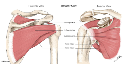

1) Rotator cuff and Proximal Biceps Tears (partial vs. full thickness, acute/traumatic vs. chronic/degenerative)

  • the rotator cuff is responsible for shoulder rotation, stabilization, and arm elevation
  • the rotator cuff holds the head of the humerus in the small shallow glenoid
  • rotator cuff muscles: supraspinatous, infraspinatous, teres minor, subscapularis
  • rotator cuff tear very common, 2 million people in US/year
  • important history in your diagnosis of rotator cuff injury: usually in dominant arm, age >40 years, pain worse at night, interferes with sleep, weakness with rotation and lifting, unable to do daily tasks (e.g. combing hair, putting on shirt)
  • Exam
  • acute traumatic usually occurs in setting of fall, trauma, significant amount of force (particularly in young patients)
  • chronic is degenerative, gradual onset, repetitive stress injury, occurs often in dominant arm, often as a result of bone spurs rubbing
    • >40% of people >65 have chronic rotator cuff tear
  • rotator cuff tears DO progress with time
    • full thickness tendon tears progress more rapidly
    • larger tears progress more quickly as well
  • Non-surgical management: activity modification, NSAID, cortisone?? (controversial, Dr. Pourtaheri doesn't recommend steroid injection for rotator cuff injury), PT helps with strength and pain, doesn't fix the tear, work on strengthening other tendons
  • All acute traumatic tears in people <60 should be fixed
  • "old tendons are not fixable" (no atrophy on MRI)
  • Shoulder arthroscopy: small incisions w/camera, nerve block for pain
  • Rotator cuff repair in correct candidates have 95% success rate (in terms of pain, function), improved shoulder strength and prevent tears from progressing
  • Post op course: 6 weeks in sling, 3 month limited lifting, PT

Many rotator cuff tears have an associated biceps tendon tear (should be repaired at the same time)

2) Impingement/Bursitis

  • inflammation of the bursa, usually due to overuse (overhead activity), sometimes trauma
  • pain exacerbated with activity, relieved with rest/NSAIDs, immobility
  • no-op treatment: activity modification, steroid injection, ice, ultrasound, PT
  • arthroscopic surgery for bursitis is generally arthroscopic bursectomy w/arthroplasty on the undersurface of the acromion 
  • rehab is faster than rotator cuff: sling x 2 weeks, PT within 2 weeks of surgery

3) Shoulder Labrum Tear

  • Labrum is fibro-cartilaginous ring that attaches to the glenoid, anchor point for gleno-humeral ligaments 
  • labrum is essential for shoulder stability in people <40
  • in people <40 tears are usually associated with trauma or dislocation event
  • in people >40, most labrum tears are physiologic and don't need treatment or surgery
  • PT is best non-operative management   
  • Sometimes surgery is indicated for people who are young and failed PT

4) Shoulder arthritis

  • >60 year old patient arthritis is a common cause of shoulder pain
    • articular cartilage thins out with time, exposed bone
  • X-ray: collapsed joint space, large bone spurs, thickening of subchondral
  • Non operative management: NSAID, PT, steroid/cortisone injection (yes, indicated)
    • PT to stretch the shoulder joint capsule (see exercises below)
  • Operative tx: shoulder arthroscopy (to release joint capsule, usually in mild to mod arthritis)) and shoulder replacement
  • Shoulder replacement 90% pain relief indicated for moderate-severe arthritis of gleno-humerus
  • there have been significant advances in shoulder replacement techniques and technology over the last 10 years
    • same day (outpatient surgery)
    • 4-6 weeks immobilized in sling, PT within a week, full recovery 6 months-1 year
  • two types of shoulder replacement: anatomic (intact rotator cuff) vs. reverse shoulder replacement (non anatomic)-- shoulder arthritis w/large rotator cuff tears
    • in reverse, ball goes on socket side of shoulder, socket on ball side of shoulder

Final pearls:

  • History and physical exam are key for assessment and diagnosis of shoulder pain
  • X-rays are still always a good idea as an initial evaluation (arthritis, calcific tendonitis, acromial bone spurs, for large rotator cuff tears for decision-making for surgery)
  • MRI definitively diagnose rotator cuff tears
  • Role for ultrasound? In patients who cannot get MRI (e.g. pacemaker), can use for shoulder injections (ultrasound guided)
  • Absolute indications for MRI in shoulder pain
    • fall/acute injury with sudden onset weakness in the arm likely has an acute rotator cuff tear(to evaluate for rotator cuff tear, which should be repaired within a couple months of injury for best outcome, time sensitivite)
    • if concern for biceps tendon "Popeye" arm (full thickness tear of biceps tendon)


AI in Radiology (Rael, 5/19/21)

Thanks to SMGR Radiologist, Dr. Jesse Rael, for a thought-provoking Grand Rounds titled AI in Radiology this week. Super interesting. 

The recording is available HERE for your viewing pleasure. 

Here are my thoughts on Dr. Rael's presentation: 

facial recognition at airport (cnn.com)

Artificial intelligence is the the theory and development of computer systems able to perform tasks that normally require human intelligence, such as visual perception, speech recognition, decision-making, and translation between languages.

AI is present in many aspects of modern life, including facial ID, social media platforms, google search, digital voice assistants (siri and alexa), smart home devices, amazon recommendations, and more.

The topic of AI feels very far from where we are in the world of safety net primary care-- where literally just getting a patient a follow up appointment with a PCP feels like moving a mountain. But I am not sure my sense is actually accurate. In fact, Dr. Manny Mendoza gave a Grand Rounds just a few months ago on AI in Primary Care. The link to that GR summary and recording is here. And the truth is, AI is already making changes in primary care: decision-making tools, benign vs. malignant lesions in dermatology, chronic disease management and more.

With regards to radiology, AI means a computer that is trained to interpret images to either rule in/out a radiographic diagnosis. This could include a wide range of diagnoses, from fractures to pulmonary emboli to cancer, and beyond.

Proponents of AI in radiology argue that radiologists are expensive, there is increasing expectations of productivity, that imaging is getting more complex, numbers of images per study are increasing, and that AI could streamline and improve that work. 

Dr. Rael doesn't think that computers are going to replace radiologists, BUT, he believes, radiologists who don't adopt the evolving technology will likely be replaced. In his words, AI could be considered equivalent to autopilot in a jet engine cabin-- the pilot is absolutely needed, particularly for complex situations, and the autopilot function is there to help him do his job better. 

Dr. Rael's hope is that AI will help radiologists to become more productive, enhance protocols, be available where radiologists are not, and more. And, as he points out, radiologists do much more than interpret images-- they are involved in clinician-to-clinician and clinician-to-patient communication, QA, education, policy making, hands on scanning, biopsies, etc.

Dr. Rael spent some time helping us to understand the concept of "deep learning" currently being used in image interpretation. He showed us how a computer can "see" images (e.g. a kidney or a brain lesion) on a screen.


There are many different current AI projects around the world: identification of pulmonary edema on CXR, white matter abnormalities in very preterm infants, PE recognition, benign vs. malignant breast lesions, abnormalities in knee MRI, and automatic scan range delimitation in Chest CT. The possibilities are endless! Dr. Rael himself is involved in a neuroradiologic project looking at image analysis for different brain lesions-- what does a glioblastoma look like? How can a computer recognize it?

And what about global health implications for providers working in places where there are not only no radiologists, but no machines to image (e.g. butterfly ultrasound with images being interpreted by offsite AI)? 

What do you think about AI in radiology and AI in general in medicine? Deeply interesting, kind of scary, and definitely cool.

My advice for this week: if you have a question about a radiographic finding, don't forget to call your friendly local radiologist-- I always feel like I understand more about a complex impression, after I have spoken to a real live human radiologist. Will that always be so?


Radiology Potpourri (Kujala, 3/17/2021)

Many thanks to Dr. Nick Kujala, Sutter Radiologist and mid-West Scrubs and hockey fan, who gave an entertaining and informative presentation this week, covering a range of topics in radiology: from the history of the first radiograph to the risk of radiation exposure to the invention of the CT scanner. The video recording is available HERE.

Here are a few summary points from his presentation:

1) Radiation Exposure: Many patients (and clinicians) have concerns about the risks of radiation exposure with imaging studies. It may be helpful to note that living on earth gives us daily background radiation exposure, and certain jobs/situations (e.g. working as flight attendant or pilot) increase the amount of that exposure over time. Of note,  exposure from one chest x-ray is the equivalent of  ~10 days of background radiation, whereas at CT of the chest is equivalent to ~2 years. 

Information on radiation exposure for patients is available at this website:  https://www.radiologyinfo.org/en/info.cfm?pg=safety-xray

Also, remember that MRI and ultrasound are alternative imaging modalities that offer ZERO radiation exposure.


2) Breastfeeding and contrast: Women who are breastfeeding can safely receive contrast (iodinated and gadolinium-based) for imaging studies without concern. The dose absorbed by an infant is exceedingly low. There is no need to pump and dump, but ultimately the decision should be left to the lactating mother.

3) ACR Appropriateness Criteria: The American College of Radiology (ACR) has an excellent, information-packed website to help clinicians make the correct choice about imaging studies. Everything you want to know about radiology imaging (indications, risks/benefits, radiation exposure, alternatives) can be found here: https://www.acr.org/Clinical-Resources/ACR-Appropriateness-Criteria

An excerpted example of these ACR criteria for abnormal uterine bleeding is pictured below.


4) MRI and Gadolinium: Gadolinium has been used as contrast in MRI for over 30 years. There is a known phenomenon of gadolinium deposition in some tissues (bone, kidney, brain); however, there has been no consistent evidence to suggest that these deposits are associated with neurotoxicity. However, as recent as 2016, some scientists have suggested a condition called Gadolinium Deposition Disease, linking these deposits with a constellation of neurological symptoms and signs.  While Dr. Kujala and the ACR  support the safety profile of gadolinium, it is not recommended during pregnancy

5) More IS Better! Give your radiologist as MUCH clinical information as possible when ordering an imaging study-- you will get more clear results back. And if you have a question, call your local radiologist sitting in his dark reading room (or at home) at x-44551.

Extras:

Roentgen's first ever radiograph of his wife's hand (1895) 


Hounsfield's invention: the CT Scanner (he shared the 1979 Nobel Prize in Medicine)


And finally, what is an Aunt Minnie? The origins of the term "Aunt Minnie" are a bit hazy, but it's believed to have been coined in the 1940s by Dr. Ben Felson, a radiologist at the University of Cincinnati. He used it to describe "a case with radiologic findings so specific and compelling that no realistic differential diagnosis exists."


Can you name the Aunt Minnie below?


Answer: Tension Pneumothorax


Diagnosis and Management of Osteoporosis (Hamann 7/23/2026)

 A recording of this presentation is available HERE .  *** Thanks so much to Dr. Kendal Hamann, SMGR Endocrinologist, for an outstanding Gra...