Showing posts with label emergency. Show all posts

Acute Liver Injury (Deis, 10/29/25)

 A recording of this presentation is available HERE.

Many thanks to Dr. Faith Deis for an awesome presentation this week on Acute Liver Injury (ALI). For those impatient folks out there, the most important take home points are: 

1) While the combination of alcohol and acetaminophen can actually be hepato-protective, a pattern of heavy ETOH use + fasting followed by moderate to high dose of APAP can be particularly hepatotoxic and make vulnerable patients more susceptible to ALI. 

2) N-Acetylcysteine (NAC) replenishes glutathione with is hepatoprotective and shows benefit for ALI of multiple etiologies (not just APAP intoxication); when in doubt and/or when in the midst of a work-up, give NAC empirically for any undifferentiated ALI, even if APAP level is normal.

A few more pearls from Dr. Deis's talk follow. . .

Acute Liver Failure= Acute liver injury (as indicated by elevation in AST/ALT) + Coagulopathy (INR>1.5) + Hepatic Encephalopathy

Much like many of us use LactMed for breastfeeding safety, there is an excellent NIH source, LiverTox, which compiles all the evidence we have on liver toxicity of medications. Just last night while precepting, a resident and I used this resource to understand whether GLP-1 medications may be implicated in rising transaminases in a patient with poorly controlled DM2. This is a great resource!


Dr. Deis introduced us to the idea of using the "R factor" to help distinguish between intrahepatic and cholestatic patterns of liver injury.

How to approach elevated liver enzymes? | AASLD

From the 2021 ACG Guidelines. You can see how both the LiverTox and the R factor are involved in your clinical decision about 1st line testing for abnormal liver enzymes:

A little physiology and pathophysiology of APAP Toxicity, which you will note involves the CYP-2E1 pathway (5-9% of hepatic metabolism of APAP) and NAPQI, which leads to hepatocyte necrosis. In APAP overdose, more of the pathway is pushed to the NAPQI pathway! This is important in consideration the mechanism of action of NAC, which actually helps to replenish glutathione, and therefore shift the ASAP metabolism pathway away from the toxic one and back to the healthy one.

Clinical Aspects - Acetaminophen Toxicity Diagnostics, LLC

Dr. Deis also shared an interesting set of studies on the interaction between APAP and Alcohol on liver metabolism. This is super interesting! It turns out that if APAP and alcohol are ingested simultaneously, alcohol's metabolism actually has a protective effect on the liver's metabolism of APAP, keeping it in the non-toxic pathway. BUT if alcohol is ingested prior to APAP administration (and particularly in the setting of prolonged fasting, which is not uncommon during an alcohol binge), then the use of APAP is pushed toward the NAPQI pathway and is more likely to be hepatotoxic. Practically speaking, then, if a patient has an alcohol binge, and then consumes even moderate doses of APAP at the tail end/after the binge, those moderate doses may lead to a disproportionately toxic impact on the liver. This means, then, that you can have acute liver injury (and even failure) from a therapeutic dose of APAP. 

What about NAC?


Turns out that NAC's protective effect on the liver extends beyond APAP ingestion. In a 2021 Metanalysis , authors found a small but stastitically significant mortality benefit in patients with non-APAP induced liver injury and improved mortality (see table below). There are actually few downsides to NAC (the only absolute contraindication is allergy to NAC itself, care with volume needed to infuse for patients for whom volume could be problematic). In sum,  American College of Gastroenterology 2024 Guidelines actually recommend NAC be administered to all patients with ALI while work-up is in progress. 


A bit on Alcoholic Hepatitis (which may be on your ddx in someone with binge drinking behaviors)
-Remember the Alcoholic hepatitis rarely has AST/ALT elevated above 400-500 and the AST/ALT ratio should be somewhere around 1.5 and the Total Bilirubin is almost always >3.

The 2024 ACG guidelines on alcohol associated liver disease have a really nice set of flowsheets (for those of you who love flowsheets) that nicely outline the evaluation of Alcoholic hepatitis and the subsequent decision tree around using (or not using) steroids, remembering that steroids have downsides (infection, GI bleed, hyperglycemia, AKI, psych) and should only be used in appropriate candidates. 
 
On a final note, we must be careful with patients with ALI who also undergo alcohol withdrawal. Phenobarbital, which has become widely used in our institution in the last year, is relatively contraindicated, as are sedating medications (e.g. benzos). That being said, alcohol withdrawal is a dangerous condition that could result in ICU transfer and/or even death, so we need to be treating them. A reminder that librium/chlordiazepoxide, which has widely fallen out of favor, may be the best option in these patients. 

So much good learning!
Thanks for making it to the end.

Dr. Deis' references:

ACG Guidelines 2024

LiverTox

AASLD

Ghosh, A., Berger, I., & Remien, C. H. (2020). The role of alcohol consumption on acetaminophen-induced liver injury: Implications from a mathematical model. Journal of Theoretical Biology, 510, 110559. https://doi.org/10.1016/j.jtbi.2020.110559 ouci.dntb.gov.ua+1

Forget, P., Wittebole, X., & Laterre, P.-F. (2009). Therapeutic dose of acetaminophen may induce fulminant hepatitis in the presence of risk factors: A report of two cases. British Journal of Anaesthesia, 103(6), 899-900. https://doi.org/10.1093/bja/aep322 OUP Academic

Ghosh, A., Berger, I., Remien, C. H., & Mubayi, A. (2020). The role of alcohol consumption on acetaminophen-induced liver injury: Implications from a mathematical model. Journal of Theoretical Biology, 510, 110559. https://doi.org/10.1016/j.jtbi.2020.110559. (Note: duplicate to #1 – keep one)

Lee, W. M., Kaplowitz, N., et al. (2020). Acute liver injury with therapeutic doses of acetaminophen (≤ 6 g/day): A prospective study. Hepatology, (in press). https://pubmed.ncbi.nlm.nih.gov/33306215/ PubMed+1

Whitcomb, D. C., & Block, G. D. (1994). Association of acetaminophen hepatotoxicity with fasting and ethanol use. JAMA, 272(23), 1845-1850. https://doi.org/10.1001/jama.272.23.1845 (from PubMed 7990219)



Chest Pain Workup (Peng, 3/13/2024)

 A recording of this presentation is found HERE. 

***

Thanks to Dr. Jonathan Peng for an excellent Grand Rounds this week on Chest Pain Workup. A recording of his presentation is above. My favorite favorite moment of the presentation was his last statement: "If it is important to the patient, it should be important to us." In other words, don't dismiss a patient's concerns. I love this mantra, and I so appreciate Dr. Peng's presentation -- he took us through a little history of medicine, some biostatistics, a bit of art history, and finally, the black box of cardiac stress testing.


Here are my notes:

Dr. Peng's  recipe for cardiac evaluation:

  1. Evaluate the patient by their baseline characteristics (i.e. pretest probability).What are a patient's risks for CAAD? We should all be  aware that increasing age increases the likelihood.
  2. Evaluate the patient with their symptoms (using risk scoring). How likely is it that the patient in front of me has chest pain that is ACS?
  3. Evaluate the patient with testing (EKG, laboratories, stress testing, etc.)


Cardiologists love acronyms and scores!

There are two main calculators that help us predict cardiovascular risk using risk factors

  1. ASCVD Risk Estimator (2019) predicts 10 year risk 
  2. PREVENT Online Calculator (2024), new from AHA, predicts 10-30 year risk, in specific populations
There is additional score, called the HEART Score (2011) that helps to predict a patient's risk of ACS during an acute event. Factors include:
  • History
  • EKG
  • Age
  • Risk Factors
  • Troponin
This HEART Score for Major Cardiac Events predicts a patient's 6-week MACE (Major Adverse Cardiac Event) risk. A score of 0-3 is low risk (1.7%), 4-6 is intermediate risk (16%) and 7-10 is high risk (50%). The authors suggested that a low HEART score excludes a short term MACE with a 98% certainty!

Once cardiologists have decided that a patient needs evaluation, they consider the following:
  • indications: why am I ordering the test?
  • contraindications: why should I NOT order the test?
  • types of test: which one to choose?
  • interpretation: what do the results mean?
Stress tests are ordered to rule out ischemia and to assess for cardiac viability, but also to assess functional capacity (eg. in valvular heart disease, hypertrophic cardiomyopathy, cardiac vs. lung dz and preop capacity if otherwise unclear), risk stratify specific patients after an acute MI (if cardiac cath not performed), and assess efficacy of medical therapy. 

Dr. Peng reminded us that many such tests are likely to be most  helpful for patients in the middle -- that is patients who have a low pretest probability will likely have normal tests, and patients with known severe CAD will have abnormal tests. 
Complications from stress tests are extremely rare, as are complications from pharmacological stress. Absolute contraindications include: severe AS, recent MI (within 2 days), unstable angina, decompensate heart failure, unstable arrhythmia, acute PE, suspected aortic dissection, inability to walk. Relative contraindications include: left main disease, moderate AS, HCM, electrolyte abnormalities, afib w/RVR, high degree AV block, and resting BP >200/110.

All stress tests involve a stress (either treadmill/exercise or pharmacologic) and follow-up imaging (EKG, echo, or nuclear med). See diagram below for the menu of stress tests:


What they are looking during the stress for something called the ischemic cascade; that is, as stress is put on the heart in the form of an exercise load, you can see a cascade of events that occur in the setting of decreasing coronary blood flow. I really LOVE this image Dr. Peng shared about how that manifests on perfusion scans, echocardiograms and EKGs, as well as symptoms. 


Exercise (i.e. treadmill testing) remains the stress test of choice -- it's cheap, you don't need special equipment, and it gives us an idea of a patient's functional capacity. It does have lower sensitivity/specificity than pharmacologic stress and there are some conditions that make a stress EKG uninterpretable (e.g. people with pacemakers, LBBB).

Pharmacologic testing options include adenosine (a vasodilator), dobutamine (a beta agonist), and lexiscan (most commonly used, shown to be safer, okay in people with BOPD).

Nuclear isotopes used in the nuclear portion of the stress test expose patients to 48 hours of high dose radiation. These types of nuc med tests can last anywhere between 2-4 hours and 2 days. Patients with higher BMI (>32-35) will often require a higher tracer dose and a longer test. 
example of nuc med imaging protocols
Stress imaging can be either via echocardiogram or nuclear med studies. Echo tends to be more specific (i.e. better for "ruling in" ischemia), nuclear med imaging tends to be more sensitives (i.e. better for "ruling out" ischemia). Other differences are seen in the table below. 


The next slide is probably my favorite from his talk, I'll call it a "Dummy's guide to stress testing". It is a flowsheet that takes us through the patient's clinical scenario and leads us down a path of which stress test to choose. Dr. Peng stressed that having good echo techs (which we do!) is an important caveat to which tests you order. 

att: Dr. Jonathan Peng, 3/13/2024

A treadmill EKG is 68% sensitive and 77% specific. In comparison, a treadmill echocardiogram has slightly increased sensitivity and specificity at 81% and 88%, respectively. A treadmill myoview has sen/spec of 87% and 70%. This makes me conclude that if we had a good echo tech to do the study, I would prefer a treadmill echo for myself (to avoid the radiation!).

What does an adequate study mean?
The results of stress tests don't always seem obvious. And this is because, you must first understand if the test was adequate before being able to interpret the results. Adequacy is defined as 1) patient being able to reach target heart rate,  (max predicted heart rates is traditionally defined as 220-age in years; the target heart rate is 85% of the max heart rate). A stress test is only negative IF the patient reached the target heart rate. 

On a related note, should we hold a beta blocker before a stress test?
It depends: for diagnosing ischemia then YES because you need to reach the target heart rate. For evaluation of medication efficacy then NO because we want to know if the meds are working correctly

It is also important to understand why a stress test was stopped. Absolute reasons for stopping include: ST elevations, BP drop >10mm hg with evidence of ischemia, mod/severe angina, dizziness/syncope, cyanosis/pallor, and sustained VT. Relative reasons for stopping the test early include: asymptomatic drop in BP, ST depressions >2mm, increasing chest pain, multiple PVCs, BP>220/115, fatigue/SOB/leg pain. 

Exercise time is prognostic! >8-9 minutes on the treadmill is a good prognostic sign. 

Of course, since we are talking cardiology, there are different scales to assess the outcomes of a treadmill tests and to use that to predict CV morbidity and mortality. These include the Duke Treadmill Criteria and the Cleveland Clinic score. The Duke score takes into account how long you were able to exercise, how bad the EKG changes, and whether or not you had chest pain during the test. The higher the number, the higher the CV risk.

High risk stress tests involve people with short time to symptom (3 minutes or less), induced hypotension, and prolonged time to recovery. The notion that having your heartrate return to baseline with relative quickness predicts the severity of your cardiac disease. For more high risk stress test outcomes, see the image below.


Unsurprisingly, exercise ability predicts prognosis -- that is, the more you can exercise (i.e. the higher METS you  achieve), the better your prognosis, regardless of your comorbidities. This is a reminder to us all to move move move our bodies as much as we possibly can! Stay in shape. 

Dr. Peng spent just a few minutes at the end talking about CT coronary angiography, a relatively new imaging study for intermediate and low-risk patients, those who are younger, have a lower BMI. It exposes people to much less radiation than a nuclear med study and decreases a need for unnecessary cardiac catheterization in the lower risk population. 

And finally, Dr. Peng ended the way I began this blog --with a plea to listen to patients: "When I was a younger clinician, I used to ignore my patients, sometimes disregard their symptoms. They would complain of pain or shortness of breath, and I would think 'oh, that isn't cardiac'. With time, I have learned to listen to my patients. If it is important to them, it should be important to us."

Amen. Dr. Peng.
Go forth and listen to your patients. 

Oncologic Emergencies (Kanaan - 9/13/23)

 A recording of this presentation can be viewed HERE.

***

Thank you to local SMGR Oncologist, Dr. Zeyad Kanaan, for an action-packed presentation this week on Oncologic Emergencies. His presentation focused on five major emergencies-- SVC syndrome, Tumor Lysis syndrome, Hypercalcemia of malignancy, Acute malignant cord compression, and Leukostasis. Sprinkled among the five oncologic emergencies was a necklace of pearls for the primary care provider caring for cancer patients. 

Here are a few general pearls:

  • Primary care providers diagnose cancers; oncologists treat them. 
    • Oncologists cannot begin treatment of any cancer without a biopsy (i.e. "tissue is the issue")
    • It can be anxiety-producing to carry a patient through the process of a cancer discovery and arranging the appropriate plan for biopsy. 
    • Oncologists can help guide us through this part, even if they are not yet managing the cancer.
  • All cancer symptoms (e.g. chemo-related nausea and vomiting) are graded 0-5 (1 is mild, 5 is most severe). 
    • Most people with Gr 1 &2 symptoms can be managed as outpatient. 
    • Patients with Grade 3 sx should be sent to the ER. 
    • Gr 4 is life-threatening (often means ICU).
  • Aggressive cancers, by definition, replicate and grow rapidly, which means they also tend to respond quickly to treatments. 
    • BUT aggressive cancers can also can come back quickly
  • All oncologic emergencies require an urgent management plan, a plan for the next few days (less urgent). 
    • Ultimate management is treatment of the underlying cancer.
SVC Syndrome
SVC syndrome is caused by obstruction of the SVC, leading to facial edema and sometimes arm edema. The most common cause of SVC syndrome is cancer (particularly lung cancer, lymphoma, germ cell tumors). But there are other less common things that can obstruct the SVC. 

Medial and perihilar masses are more likely to be squamous cell cancer and small cell lung cancers.

If you see "central necrosis" on imaging it means that the cancer is very aggressive and is growing too fast for its own blood supply.

As mentioned above, SVC Syndrome is graded 0-5. Grade 3 symptoms require ER/hospital management and definitive treatment of the underlying cancer, which means urgent initiation of chemotherapy.
Grade 4 requires a stent (rarely done).

SVC syndrome, regardless of Grade, is a bad prognostic sign. 

Tumor Lysis Syndrome (TLS)
TLS is an oncologic emergency caused by massive tumor cell lysis with the release of large amounts of potassium, phosphate, and nucleic acids into the systemic circulation. TLS is more common in solid tumors. TLS can lead to AKI, cardiac arrythmias and even seizures. TLS can be diagnosed either clinically or via lab tests (lab abnormalities: hyperkalemia, hyperphosphatemia, hypocalcemia, and hyperuricemia). TLS can occur spontaneously with aggressive cancers and/or can also occur with initiation of chemotherapy.


Treatment of TLS:
1) Aggressive Hydration (1-2L bolus, followed by 200-300ml/hr crystalloids)
2) Urine alkalization (via bicarbonate drip)
3) Allopurinol -- allopurinol is a preventive drug for TLS, does nothing to an already elevated uric acid and cannot help urgently. Given PO. Dosed 100mg/m2 or 10mg/kg/day (divided q8h). Often started in solid tumors prior to chemo initiation.  Dose needs to be reduced by 50% with renal failure.
4) Rasburicase -- considered a rescue agent, very potent cleavage of uric acid, $$$, very effective within 30 minutes. Usually requires only one dose (3mg, which is half of the manufacturer recommended dose), check uric acid daily. Can repeat but hardly ever necessary.

Both allopurinol and rasburicase should be started together in cases of TLS. Of note, both allopurinol and rasburicase can (and should) be given prophylactically in cases where TLS is highly anticipated. 

Hypercalcemia of Malignancy
"moans, bones, and psychic overtones"
Lytic cancers that metastasize to bone are most likely to cause hypercalcemia. These include, most commonly breast cancer, multiple myeloma, lymphomas, and leukemias. Prostate cancer, e.g. while often spreading to bones, is less likely to be lytic.
Treatment of Hypercalcemia:
1) Aggressive hydration
2) Bisphosphonates (+Denosumab) take a little longer than calcitonin. Works in ~2 days. 
3) Calcitonin: is the acute treatment of choice, will drop the calcium quickly, but Ca will rise again quickly if otherwise left untreated

Once Ca starts dropping, goal is to treat the underlying cause.

Don't forget to check labs to be sure it's hypercalcemia of malignancy. A low intact PTH and elevated PTHrP is most consistent with malignant hypercalcemia. HyperCa in setting of elevated vitamin D levels, more often seen in lymphoma, is more likely to respond to steroids.

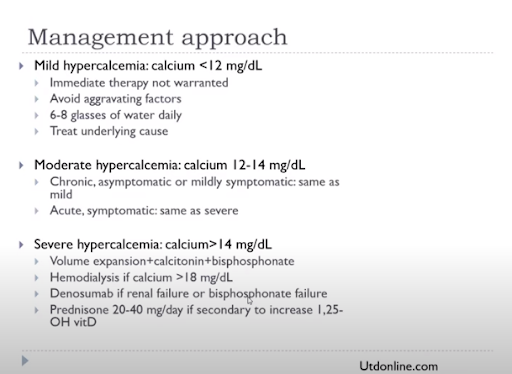
Mild hyper Ca (<12): oral hydration, treat the underlying cause
Mod hyper Ca (12-14): need IV hydration
Severe hyper Ca (>14): IVF, calcitonin, bisphosphonates, Hemodialysis can be helpful if Ca>18, desnosumab if renal failure. prednisone if elevated Vitamin D

In the end, once again you must treat the underlying cause. 

Acute Malignant Cord Compression
This is a true oncologic emergency.

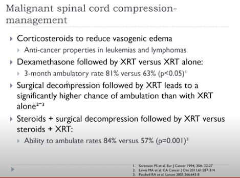
The thoracic vertebrae is the most common spinal place for cancer to go (lumber spine is the next most common). The cancer spot may actually be very small, but it is the edema leading to the cord compression and symptoms of pain and weakness. A patient's pre-intervention neurologic status influences a patient's chance of regaining function

Hours can matter in terms of neurological preservation, so do not be afraid to start steroids. Primary care and ED's job is to 1) start steroids -- dexamethasone 4mg q6 hours (total 16 mg/day) and 2) call the neurosurgeon. 

The vasogenic edema will respond quickly to the steroids. 

Leukostasis = hyperviscosity (of Waldenstrom)
Most common symptoms: bleeding, blurred vision, headache, vertigo
Treatment is IVF to decrease viscosity
Check serum viscosity (>4 is significant)
Check IgM level 
Plasmapheresis is very effective
Local patients with acute leukemia are admitted for urgent chemo (either to Alta Bates or Sacramento)
Goal is to treat leukostasis (WBC<100K), end organ effects (e.g. CAD) and leukemia all at once

Pediatric Trauma (Bellman, 12/8/2021)

 A big thanks to Dr. Lilly Bellman of CPMC Pediatric Emergency Medicine for her presentation on Pediatric Trauma this week.

A recording of her presentation is available HERE

We learned that trauma in children is different: remember KIDS ARE SQUISHY
    *many have internal injuries without much sign on the outside
    *children have bigger heads proportionally
    *their bodies are more flexible and their bones are less calcified
    *their abdominal organs are less protected and relatively larger, thus more susceptible to trauma

Clinical decision tools can help us determine risk and evaluation. They help us identify children that are at lower risk and in whom we can avoid imaging.
  • Head CT has 1:6,000 lifetime risk of fatal cancer
  • Abdominal CT has 1:1,000 lifetime risk of fatal cancer
The mechanism matters, for example in an MVA, how fast? restrained? rollover? need for extrication? Or a fall - from what height? onto what surface?
What is your clinical evaluation - are they acting normally? ambulatory? LOC? witnesses? Intoxicated? and of course - last Td? (if they are 10+ may not be protected)

Decision Tools: 

Nexus Criteria for neck injuries
In addition to Nexus, get a Neck CT if trauma + torticollis. Get Neck CT or XR if midline tenderness. Get MRI if abnormal neuro findings
Inline image

Blunt abdominal trauma algorithm: 
Inline image

and there are several other useful decision tools...

When to get imaging in children with head trauma 


Finally, don't forget about NAT (non-accidental trauma) any time there's a history and/or mechanism inconsistent with injuries or the child's development.

If they can't cruise, they can't bruise....

  • Sentinel injuries include: bruises (trunk, ears, neck) <4 yo, oral injuries in infants, or patterned bruises or burns
  • Evaluation: skeletal survey (<2 yo), screening labs for occult abdominal trauma (LFTs, lipase), have a low threshold for head CT, and consult CPS. 
  • Protocolized systems for NAT screening are helpful to reduce bias - remember to check your biases along with your suspicions!

What Every Health Provider Needs to Know about Drowning (Hoffman, 11/3/2021)

 Thank you to Dr. Ben Hoffman who gave a profoundly moving talk on Drowning Prevention in Children. What an honor to host the national expert on accident prevention!

A recording of the excellent presentation is HERE. 

I consider this presentation a MUST for all of us that care for children. This is PRACTICE CHANGING.

Here are our notes:


Drowning is the leading cause of unintentional injuries in children 1-18 from data collected between 2009-2018-- that’s over 9,000 children. 

  • Imagine 9 school buses of children-- 72 kiddos in each bus-- that die every year from drowning.
  • Drowning is the single leading cause of death in children ages 1-4, and the 2nd leading cause in children 15-19.
  • We must remember the BIPOC community who suffer disproportionately from incidents of drowning.
    • we can trace this reality back to systemic racism and lack of access to pools, swimming lessons, etc.

 PREVENTION: the AAP has created both a toolkit https://www.aap.org/drowning and a policy (attached) to help providers educate family’s on drowning prevention. The toolkit has both general information and patient handouts and posters for your office.

https://www.aap.org/drowning

The bottom line: LAYERS OF PROTECTION to prevent drownings, and we should focus particularly on new parents of children <4, teens, BIPOC families, and children with disabilities and epilepsy.

                *infants: never leave unsupervised – even a second- in water.

                *toddlers: their curiosity is dangerous. Never leave a toddler unsupervised around any water. They can get into tubs, toilets, wading pools. Lock or empty these when not in use.

                *swimming lessons: no evidence that they protect infants, but there IS evidence that they work for children 1-4 yrs old and shows a significant reduction in drownings.

                *water competence: we should teach our families that learning to swim is a life skill. Many BIPOC parents were never taught to swim so consider the water dangerous.

                *erect barriers: particularly pools – 70% of pool drownings are when it’s not “swim time”. Pool fences that have 4 sides with a locking gate reduce drownings by 50%.

                *supervision: constant, close and capable supervision-at arm’s length if a child can’t swim competently. Don’t rely on lifeguards – children still drown in their presence.

*life jackets: only coast guard approved life jackets are appropriate (and a must when our families visit the Russian River or the Sonoma Coast). Never rely on anything inflatable. The coast guard approved life jackets are more expensive than the inflatables.

*advocacy: some cities have life jacket loaner programs. Dr Hoffman will be glad to speak to anyone who is interested in starting a program locally (for example Spring Lake loans them with boat rentals).


Calm in the Chaos: An Approach to Rapid Responses (Bamidele, 7/28/2021)

Kudos to Dr. Stephanie Bamidele, who delivered our very first R3 Grand Rounds of the Academic year. Her topic? Calm in the Chaos: An Approach to Rapid Responses.

A recording of her excellent presentation is available HERE for your viewing. 

And here are my notes:

Anyone who has worked in a hospital knows that rapid responses occur frequently in the hospital. But it hasn't always been that way! The concept of Rapid Response Teams emerged in the 1990s, modelled after Code Teams (which originated in the 1960s), as a standardized group of people expected to respond to a call for help before a patient requires a Code Team's assistance. The RRT goal is to intervene and respond to prevent major adverse events. And then make systems changes that allow for the next response to be even more effective. 

Anyone can activate an RRT.

RRT may be activated for a number of reasons: vital sign abnormalities, nurse or family detection of clinical deterioration, a change in mental status, and more.

Specific clinical criteria may be used to designate a RR present minutes to hours before a serious adverse event:
  • HR <40 or >130
  • SBP <90
  • Respiratory Rate <8 or >28
  • Oxygen saturation <90%
  • Change in level of consciousness
  • Change in urine output (<100ml/4 hours)
Like a Code, any Rapid Response starts with the basics: A (airway), B (breathing), C (circulation) and goes from there. See these images for a very clear description of events that should occur in the first five minutes of a RR: 

A

  • AIRWAY

  • Assistance (call for help)

  • Activate RRT

  • Annunciate when providing key information

  • Acquire Data

  • Attend to patient until RRT arrives

  • Access (IV)

  • Assist as directed by team leader

B

  • BREATHING

  • Bed (away from wall)

  • Backboard if pulseless for CPR

  • Blood glucose if altered mental status


C

  • CIRCULATION: check pulse and BP

  • CPR

  • Crash Cart

  • Connect IVF

  • Clear the Room

  • Communicate to RRT

D

  • Defibrillate if Vfib or pVT

  • Document vitals at time RRT was called

E

  • Explain (S-BAR) to team leader.

    • Situation, Background, Assessment/Action, Recommendation/Report


As in all parts of life, communication is everything! 

Dr. Bamidele reminded us that a standard way of communicating (in this case, following the hospital ISBAR model) is the best way to go. Doing so, ensures that all people present at a RR, have a clear understanding of what is happening and what is being recommended.
 
Okay, doctors, so, you are the leaders of the RRT, right? What do you actually do then to be an effective leader? Here are key steps, as outlined by Dr. Bamidele:

  1. Identify team members and roles: who is the leader? what are the roles? Does everyone need to be here. Start with a simple, "I am Dr. ____, and I am leading this RR"
  2. Crowd control: limit to 8-9 people max, including the leader (doctor), ICU charge RN, bedside/flor RN, respiratory therapist, and any necessary techs (lab, xray, etc). Send everyone else back to work.
  3. Set a calm and assertive and inclusive tone. Be respectful, willing to ask for help.
  4. Promote a flattened hierarchy: think aloud, step back, voice specific findings (e.g. glucose, x ray findings, response to narcan_, ask for suggestions and/or feedback
  5. Gather info via parallel processing (this is hard!): visual assessment, forming a plan, proposing an intervention
  6. Have situational awareness: don't miss the forest through the trees, integrate evolving information in real time, reinforce plan, gather suggestions
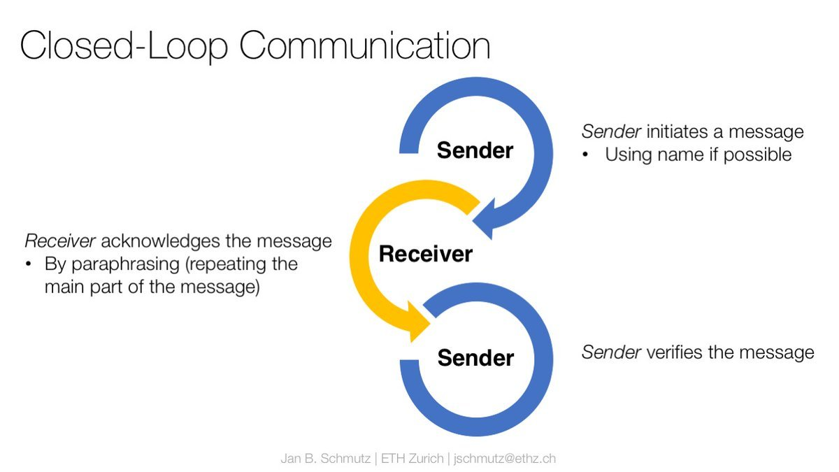
  7. Use closed loop communication: call out--> check back--> verification

Dr. Bamidele also gave us specific advice about the role of the Family Liaison during a rapid response or a Code event. There is mixed data on the harm/benefit of a family's presence at the bedside during these acute events, and so it is generally recommended that staff give the family the option of being present or leaving (without judgement). If the family is present, a designated staff person (RN, junior resident, etc) should be designated as the Family Liaison. That person should introduce themself, explain what is going on, stay by the family member's side (regardless of whether they stay or go), and provide that person an opportunity to ask questions. 

The aftermath
Once the Rapid response is done, it is super important to debrief, says Dr. Bamidele-- for two main reasons: first, because a Rapid Response often occurs in intense situations that may have a lasting impact on the team and that may require emotional processing and reflection. Second, reflecting on the process will hopefully allow you to do a better job next time. 

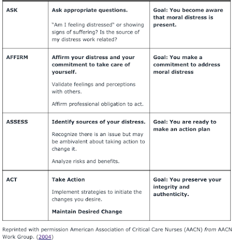
To address possible moral distress occurring in this context, consider the model of the 4As from the American Association of Critical Care Nurses (in graphic below)

Dermatologic Emergencies and their Mimics (Sugarman, 9/30/2020)

 Thanks to Dr. Jeff Sugarman for an excellent Grand Rounds this week on Dermatologic Emergencies and their Mimics. Dr. Sugarman's presentations are always replete with photos ("A picture is worth a thousand words" for sure) and probing questions, so this post will be filled with the same. Answers can be found at the very end of the post in the COMMENTS section. Don't cheat; take the quiz and use the HINTS not only to guide you to your answers, but also to enhance your understanding of the condition. 

First, when should you worry about possible dermatologic emergencies?

  • Age (newborn and young infants)
  • High fever, toxicity
  • Morphology: particularly blistering, mucosal involvement, hemorrhage
  • Specific medications: anticonvulsants, antibiotics, NSAIDs
Remember the presentation was on dermatologic emergencies and their mimics. This summary/quiz contains both derm emergencies and benign derm conditions that look pretty similar, so keep serious and not serious things on your differential. 

1) What is this rash?

Hint #1: it's really common (especially in children and people with atopy)
Hint #2: morphology includes wheals, annular, dusky centers
Hint #3: time course is VERY helpful: lesions tend to self resolve in hours, disappear and reappear in different locations
Hint #4: triggers include allergy, autoimmunity, drugs (9%), URI (40%), and idiopathic (50%)
Hint #4: Treatment: non-sedating antihistamine (fexofenadine, cetirizine) in day, sedating antihistamine at night (hydroxyzine, diphenhydramine). 
Hint #5: Prednisone is NOT rx of choice-- it works really well, and then the rash will come right back as soon as it's stopped.

2) What is this rash?
Hint #1: Looks a lot like the first rash but is different.
Hint#2: Rash morphology includes target lesions with 3 zones: dusky center, pale edematous ring, peripheral erythematous margin
Hint #3: lesions are discrete, they do NOT coalesce
Hint #4: usually pts have no systemic symptoms

3) What is this rash?

Hint#1: Presents as dusky urticaria PLUS edema, +/- fever, malaise and arthritis (7-21 days after exposure)
Hint #2: Lesions last longer than true urticaria
Hint #3: This is a type III hypersensitivity reaction (immune complexes)
Hint #4: Triggers include meds (cefaclor, PCN, anti-cancer, anti-depressants, anticonvulsants, htn meds, anti-inflammatory meds), biologic agents (rituximab, infliximab, efalizumab), infections (strep, HBC, HCV)

4) What is this rash?


Hint #1: This is a form of leukocytoclastic vasculitis in children age <2 years old
Hint #2: Presents as purpuric edematous plaques with target-like pattern, often described as "cockade or rosette"
Hint #3: This includes dramatic skin findings, but children paradoxically are not really toxic
Hint #4: Rash tends to spare the trunk
Hint #5: Lesions resolve spontaneously in 1-3 weeks
Hint #6: No labs or treatment needed.

5) What is this rash?

Hint#1: This rash may accompany pneumonia by this same organism
Hint #2: Tends to be mucosal predominant (94% oral, 82% ocular, 63% GU) and is mucosal alone in 34% of cases
Hint #3: Mean age is 12 years old
Hint#4: Most patients (81%) have no long term sequelae
Hint #5: I never heard of this before this lecture by Dr. Sugarman

6) What is this rash?


Hint#1: Severe life-threatening mucocutaneous disease involving systemic signs: fever, respiratory symptoms
Hint #2: It's a clinical syndrome, there is no definitive diagnostic test
Hint #3: Always involves at least 2 mucous membranes (mouth, eyes, urethra)
Hint #4: Causes in kids include meds (antibiotics, antiepileptics, chemotherapy), as well as HSV, mycoplasma and some undetermined causes

7) What is this rash?

Hint #1: begins as localized often occult infection (can be in the nasopharynx, perioral, conjunctiva, umbilicus, paronychia, urine, middle ear)
Hint #2: Progresses to generalized erythema and skin fragility
Hint #3: Empiric treatment is anti-staph antibiotics (cover for MRSA)
Hint #4: Peeling is NOT full thickness

8) What is this rash?
Hint #1: Most common cause of nonsexually related acute genital ulcers (NRAGU)
Hint #2: Ulcers are painful, well demarcated, shallow erosions on a clean fibrinous base
Hint #3: Self-limiting condition, usually resolving spontaneously within 2-6 weeks

9) What is this rash?

Hint #1: thick crusts, thick walled pustules are common
Hint #2: facial, periorbital involvement common 
Hint #3: fever and pain are common
Hint #4: You should culture this
Hint #5: Keflex and mid-potency steroid for body (TAC) and low potency steroid (2.5% hydrocortisone) are both indicated
Hint #6: Bleach baths (1/2 cup in full bath, 1/4 cup in 1/2 bath) may also be indicated

10) What is this rash?

Hint #1: People with eczema are particularly vulnerable to this condition due to their disruption of epidermal barrier
Hint #2: Fever, malaise, and lymphadenopathy may be present
Hint#3: This is PAINful
Hint #4: Morphology includes "monomorphous punched out erosions" (especially if you look at the periphery of this rash)
Hint #5: Lesions favor areas of active dermatitis, particularly head, neck and trunk
Hint #4: There is often a delay in diagnosis of this condition
Hint#5: Viral culture/PCR will give you the answer
Hint #6: Prompt high dose acyclovir is treatment of choice (PO for mild, IV for mod/severe)

Understanding Hospice Care (Saeed, 9/30/26)

A recording of this presentation is available  HERE .