RSV Vaccines (Teng - 9/20/23)

 A recording of this presentation can be viewed HERE.

***

Many thanks to Dr. Angelica Teng for a breaking-news Grand Rounds presentation on the brand new RSV vaccines that are rolling out just in time for the 2023-24 RSV season. A recording  of her presentation is available at the link above. 

My written notes below:

RSV, as you may already know, is an RNA virus that infects the lungs and leads to lower respiratory tract infections (LRTIs). It is the most common cause of LRTIs in infants and toddlers, leading to 2.1 million clinic visits per year in children <5 years old. RSV has less impact on adults, but does lead to 60-160K hospitalizations per year in adults >65.

Here is the link to an excellent video demonstrating classic findings in RSV bronchiolitis: https://www.youtube.com/watch?v=oOrty5PfhTY

RSV tends to be seasonal, with a typical RSV illnesses  starting in early fall (usually November in CA), peaking in winter (February), and then tapering off in early spring. It spreads mostly via droplets (cough) and can live up to six hours on hard surfaces
source: CDC

Here is the good news!

There are THREE new RSV vaccines approved in 2023
Abrysvo 
Arexvy
Nirsevimab 

There are THREE groups of patients we should consider for the new RSV vaccinations
1) Adults over 60 (Abrysvo or Arexvy)
2) Infants <8 months (Nirsevimab)
3) Pregnant women between 32-36 weeks EGA (Abrysvo)

Adults > 60: Abrysvo and Arexvy are recombinant vaccinations, each made slightly differently with inactivated proteins. Both were FDA approved in May 2023 for adults over 60. In RCTs, both vaccines were found to reduce the risk of RSV-lower respiratory tract infection by 82-89% in older adults in the first year after receiving the vaccine.  They are similar to one another, except for the inclusion of an adjuvant in Abrysvo formulation.

Infants < 8 months: In July 2023, the FDA approved a new monoclonal antibody called Nirsevimab for infants ages 1 week to 8 months during RSV season. Monoclonal antibody therapy is not technically a vaccine but can provide an infusion of prefabricated antibodies, giving temporary protection for up to five months to newly developing pediatric immune systems. In a clinical trial, nirsevimab was ~77effective against both hospitalizations and cases of RSV requiring a doctor’s visit. The CDC is  recommending that all infants <8 months during RSV season  receive Nirsevimab and additionally children ages 8-24 months with high risk conditions (e.g. prematurity, congenital heart disease, cystic fibrosis, neuromuscular disorders).

Pregnant patients 32-36 weeks: Finally, in August of 2023, the FDA approved Abrysvo as the first-ever maternal vaccine intended to help protect newborns against RSV. It was studied in late third trimester (32-36 week EGA) with the idea that protective IgG from the RSV vaccine moves across the placenta and grant protection for newborn infants.  The vaccine was found to be 82% effective at protecting infants from severe illness during the three months after birth and waned to 69effective over six months during a double-blind study. Of note, ACOG has not yet put out a formal statement about this vaccine. This is expected by late October.

A few key notes to consider:
1) Unlike a more traditional age-based recommendation for vaccination in adults >60, the CDC recommends for Abrexvy and Abrysvo that the decision to vaccinate be a shared decision between the physician and the patient. Consideration should be based on risk, and certain conditions are associated with higher risk of severe disease (e.g. lung disease, renal disease, nursing home and long term care facility residency). 
2) The RSV for pregnant woman is in no way intended to protect the mom from RSV infection and complications. It is purely meant for the infant.
3) There was a non-statistically significant outcome of low birth weight in women who received vaccination in the third trimester.
4) While there is no official statement, it is believed that guidelines will ultimately recommend that parents choose between the maternal vaccine at 32-36 weeks and the nirsevimab for infants. There is no current belief that children will need both. But this could change.
5) The only absolute contraindication to vaccination with Abrexvy or Abrysvo is anaphylaxis to any of its components.
6) It is okay to administer the RSV vaccine at the same time as either the flu or the COVID vaccine (local recommendation is to do 2 but not all 3 at the same time)
7) It is okay to administer the RSV vaccine when someone has mild cold symptoms
8) The monoclonal antibody nirsevimab is though to have a 5 month protective duration
9) It is unclear when/whether nirsevimab will replace palivizumab (synagis), the old very expensive monthly monoclonal ab that is currently used to treat high risk preemie babies during RSV season. It is likely that a single dose of nirsevimab will be more cost effective.
summary chart, source: A Teng


Oncologic Emergencies (Kanaan - 9/13/23)

 A recording of this presentation can be viewed HERE.

***

Thank you to local SMGR Oncologist, Dr. Zeyad Kanaan, for an action-packed presentation this week on Oncologic Emergencies. His presentation focused on five major emergencies-- SVC syndrome, Tumor Lysis syndrome, Hypercalcemia of malignancy, Acute malignant cord compression, and Leukostasis. Sprinkled among the five oncologic emergencies was a necklace of pearls for the primary care provider caring for cancer patients. 

Here are a few general pearls:

  • Primary care providers diagnose cancers; oncologists treat them. 
    • Oncologists cannot begin treatment of any cancer without a biopsy (i.e. "tissue is the issue")
    • It can be anxiety-producing to carry a patient through the process of a cancer discovery and arranging the appropriate plan for biopsy. 
    • Oncologists can help guide us through this part, even if they are not yet managing the cancer.
  • All cancer symptoms (e.g. chemo-related nausea and vomiting) are graded 0-5 (1 is mild, 5 is most severe). 
    • Most people with Gr 1 &2 symptoms can be managed as outpatient. 
    • Patients with Grade 3 sx should be sent to the ER. 
    • Gr 4 is life-threatening (often means ICU).
  • Aggressive cancers, by definition, replicate and grow rapidly, which means they also tend to respond quickly to treatments. 
    • BUT aggressive cancers can also can come back quickly
  • All oncologic emergencies require an urgent management plan, a plan for the next few days (less urgent). 
    • Ultimate management is treatment of the underlying cancer.
SVC Syndrome
SVC syndrome is caused by obstruction of the SVC, leading to facial edema and sometimes arm edema. The most common cause of SVC syndrome is cancer (particularly lung cancer, lymphoma, germ cell tumors). But there are other less common things that can obstruct the SVC. 

Medial and perihilar masses are more likely to be squamous cell cancer and small cell lung cancers.

If you see "central necrosis" on imaging it means that the cancer is very aggressive and is growing too fast for its own blood supply.

As mentioned above, SVC Syndrome is graded 0-5. Grade 3 symptoms require ER/hospital management and definitive treatment of the underlying cancer, which means urgent initiation of chemotherapy.
Grade 4 requires a stent (rarely done).

SVC syndrome, regardless of Grade, is a bad prognostic sign. 

Tumor Lysis Syndrome (TLS)
TLS is an oncologic emergency caused by massive tumor cell lysis with the release of large amounts of potassium, phosphate, and nucleic acids into the systemic circulation. TLS is more common in solid tumors. TLS can lead to AKI, cardiac arrythmias and even seizures. TLS can be diagnosed either clinically or via lab tests (lab abnormalities: hyperkalemia, hyperphosphatemia, hypocalcemia, and hyperuricemia). TLS can occur spontaneously with aggressive cancers and/or can also occur with initiation of chemotherapy.


Treatment of TLS:
1) Aggressive Hydration (1-2L bolus, followed by 200-300ml/hr crystalloids)
2) Urine alkalization (via bicarbonate drip)
3) Allopurinol -- allopurinol is a preventive drug for TLS, does nothing to an already elevated uric acid and cannot help urgently. Given PO. Dosed 100mg/m2 or 10mg/kg/day (divided q8h). Often started in solid tumors prior to chemo initiation.  Dose needs to be reduced by 50% with renal failure.
4) Rasburicase -- considered a rescue agent, very potent cleavage of uric acid, $$$, very effective within 30 minutes. Usually requires only one dose (3mg, which is half of the manufacturer recommended dose), check uric acid daily. Can repeat but hardly ever necessary.

Both allopurinol and rasburicase should be started together in cases of TLS. Of note, both allopurinol and rasburicase can (and should) be given prophylactically in cases where TLS is highly anticipated. 

Hypercalcemia of Malignancy
"moans, bones, and psychic overtones"
Lytic cancers that metastasize to bone are most likely to cause hypercalcemia. These include, most commonly breast cancer, multiple myeloma, lymphomas, and leukemias. Prostate cancer, e.g. while often spreading to bones, is less likely to be lytic.
Treatment of Hypercalcemia:
1) Aggressive hydration
2) Bisphosphonates (+Denosumab) take a little longer than calcitonin. Works in ~2 days. 
3) Calcitonin: is the acute treatment of choice, will drop the calcium quickly, but Ca will rise again quickly if otherwise left untreated

Once Ca starts dropping, goal is to treat the underlying cause.

Don't forget to check labs to be sure it's hypercalcemia of malignancy. A low intact PTH and elevated PTHrP is most consistent with malignant hypercalcemia. HyperCa in setting of elevated vitamin D levels, more often seen in lymphoma, is more likely to respond to steroids.

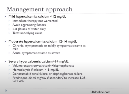
Mild hyper Ca (<12): oral hydration, treat the underlying cause
Mod hyper Ca (12-14): need IV hydration
Severe hyper Ca (>14): IVF, calcitonin, bisphosphonates, Hemodialysis can be helpful if Ca>18, desnosumab if renal failure. prednisone if elevated Vitamin D

In the end, once again you must treat the underlying cause. 

Acute Malignant Cord Compression
This is a true oncologic emergency.

The thoracic vertebrae is the most common spinal place for cancer to go (lumber spine is the next most common). The cancer spot may actually be very small, but it is the edema leading to the cord compression and symptoms of pain and weakness. A patient's pre-intervention neurologic status influences a patient's chance of regaining function

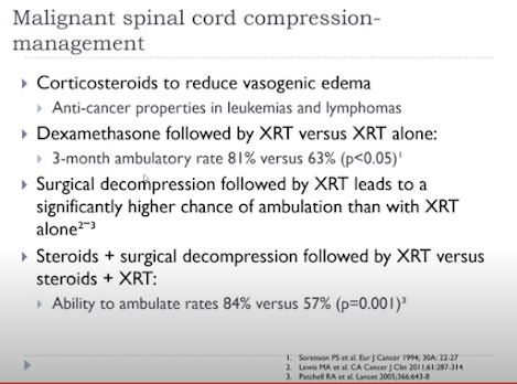
Hours can matter in terms of neurological preservation, so do not be afraid to start steroids. Primary care and ED's job is to 1) start steroids -- dexamethasone 4mg q6 hours (total 16 mg/day) and 2) call the neurosurgeon. 

The vasogenic edema will respond quickly to the steroids. 

Leukostasis = hyperviscosity (of Waldenstrom)
Most common symptoms: bleeding, blurred vision, headache, vertigo
Treatment is IVF to decrease viscosity
Check serum viscosity (>4 is significant)
Check IgM level 
Plasmapheresis is very effective
Local patients with acute leukemia are admitted for urgent chemo (either to Alta Bates or Sacramento)
Goal is to treat leukostasis (WBC<100K), end organ effects (e.g. CAD) and leukemia all at once

Thriving or Surviving: The Connection Between Chronic Stress, Chronic Disease, and Social Determinants of Health (Deol - 9/6/23)

 A recording of this presentation can be viewed HERE

***

Thank you to Dr. Navi Deol, PGY3, who gave an excellent presentation this week titled Thriving or Surviving: the intersection between chronic stress, chronic disease, and the social determinants of health. Each of the items in the subtitle is a HUGE topic, and Dr. Deol was able to weave them together beautifully and powerfully. I encourage you to watch yourself.

Image from https://www.glasbergen.com/stress-management-cartoons/cartoons/page/3

If you prefer the written word, see my notes:

  • stress: how certain stimuli (stressors) affect a person's mind, body and spirit
  • stress response: how our body reacts normal, a normal physiologic and psychologic response to stressors
  • some stressors evoke positive emotions and can be beneficial (eustress); some evoke negative emotions and cause problems (distress)
A certain amount of stress is important and necessary to generate optimum productivity and performance, but too much stress can lead to anxiety, overload, and burnout (see image below of the Yerke's-Dodson Law).
Yerke's-Dodson Law, image fromhttps://stock.adobe.com/

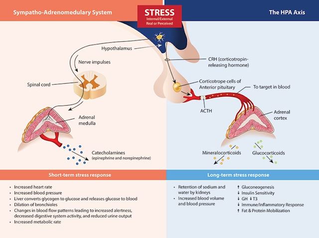
Stress causes physiologic changes in our bodies. We are all familiar with the autonomic nervous system, which responds to acute stress with the sympathetic "fight or flight" and the balancing parasympathetic "rest and digest", but what happens when the stress response is constantly being activated? 

Image from: https://www.backtothebooknutrition.com/adrenal-fatigue-hpa-axis-dysregulation/


The answer is that our long-term stress response leads to a cascade of responses that make us more vulnerable to chronic diseases.

A 2019 study published in the Journal of ACC looked at the link between SES factors such as low income and higher crime on MACEs (cardiac death, myocardial infarction, unstable angina, cerebrovascular accident, peripheral artery disease with revascularization, or heart failure). This study suggests that a biological pathway contributes to this link, involving, in series, higher amygdala activation, increased activation of the bone marrow (with release of inflammatory cells), which in turn leads to increased atherosclerotic inflammation and its atherothrombotic manifestations


Patients were categorized according to quartiles of their neighborhood median income and neighborhood crime rates.  Amygdalar activity (A) and arterial inflammation (B) were lower as neighborhood median income increased. Amygdalar activity was higher (C) and arterial inflammation trended toward an increase (D) as neighborhood crime rate increased. One image from that study is seen below. For more information, click the link above or see the study link below the image.

https://www.jacc.org/doi/10.1016/j.jacc.2019.04.042


Chronic diseases are non-communicable illnesses that persist for long periods of time and result from a combination of genetic, environmental, and psychological factors. These include cardiovascular disease (e.g. hypertension, coronary artery disease, and strokes), metabolic disorders (e.g. type 2 diabetes and obesity), mental health issues (e.g. depression, generalized anxiety disorder), and substance use disorders.

We know that inflammatory cascades play an important (and damaging role) in the onset and progression of chronic disease. While acute inflammation is technically "good" for us because it cleans up disease states in our body, chronic inflammation is bad bad bad.

Financial stress

Are you aware that money (finances and inflation) is a tremendous source of chronic stress for a shocking number of US adults. A 2022 survey found that 57% of US adults reported not having enough money to pay for essential items; 43% reported that they are not saving enough, and 56% had to make different choices due to their lack of money. 

Violent stress

Mass shootings, gun violence, and crime are also sources of tremendous chronic stress, particularly for people living in poverty and disproportionately for BIPOC people. 

Social Determinants

This leads us directly into a discussion of the importance of the Social determinants of health (SDOH), the conditions into which individuals are born, grow, live, work and age. There is an unsurprising link between chronic stress and the SDOH. These include your neighborhood and built environment, healthcare, education, economic stability, and social and community context. 

image from Healthy People 2030

If this is news to you, check out this video about how zip codes influences an individual's health: A Tale of Two Zip codes.

And for something even closer to home, check our our local Sonoma County data on how the SDOH vary based on zip code in the report titled A Portrait of Sonoma County 2021 Update, available here:  https://upstreaminvestments.org/impact-make-a-change/portrait-of-sonoma-county

https://upstreaminvestments.org/impact-make-a-change/portrait-of-sonoma-county


What can we do about all this stress?
Dr. Deol encouraged us to take a deep breath and realize that we cannot tackle these complex issues alone. First, we must acknowledge the deep-rooted history of structural and systemic racism, oppression, and discrimination that have led to health inequities that require interventions at multiple levels to reduce disparities. 

When caring for individuals, we should be careful about using the term "non-compliance" and better recognize the daily barriers our patients face due to their own SDOH. In our communities, we should be screening for SDOH and get to know and refer to appropriate community services. And at the state level, she encouraged us to support CAFP Bill AB85, which requires SDOH screening and provides resources and education for providers in referring to community health workers. 

More information for AB85 can be found HERE


Eating Disorders in Teens (Buckelew - 8/30/23)

A recording of this presentation can be viewed HERE.

Thank you to Dr. Sara Buckelew, Director of the UCSF Eating Disorder Program, who gave an excellent presentation this week on Eating Disorders in Adolescents.  As usual, a recording of the presentation is available at the link above.

Below, my notes:

Anorexia Nervosa is the most lethal of all DSM-5 diagnoses. This is a diagnoses found in all sizes and weights. Denial of symptoms and ambivalence for change are often part of the disease. Clinicians must be aware of our weight bias and the sigma associated with talking about weight. 

There are a range of eating disorders (ED), including:

  • Anorexia (including atypical anorexia, in which patient has all symptoms of anorexia but is not underweight)
  • Bulimia
  • Binge eating
  • Avoidant/restrictive food intake (ARFID)
Without question, the COVID Pandemic exacerbated eating disorders. It was a perfect storm of increased risk (increased media, disruption of school and life routines, and social isolation) and lower protective factors and access to care (including increased food insecurity, challenges to seeing primary care, etc.). This storm then led to a huge treatment bottle neck with long wait times and patients presenting increasingly ill to care.

Key Point: Early intervention is important in ED, as a shorter duration of illness is associated with improved outcomes. 

The SCOFF Questionnaire is a screening instrument designed to suggest "likely cases". A positive SCOFF does not give an adolescent a diagnosis of ED, but if positive, suspicion for ED should be elevated and early intervention should be considered

S "Do you make yourself SICK because you feel uncomfortably full?"
C "Do you worry you have lost CONTROL  over how much you eat?"
O "Have you recently lost ONE stone (15 pounds) in a 3 month period?"
F "Do you consider your self FAT?"
F "Would you say that FOOD dominates your life?"

Answering YES to at least 2 of the above is a positive SCOFF screen

Key Point: Avoidant Restrictive Food Intake Disorder (ARFID), new in the DSM-5 about 10 years ago is a more heterozygous group of ED in which there is no body image component to the restriction.

The diagnosis of ARFID concerning weight patterns, concerning behaviors: avoidance of meals, excessive exercise. The question that excludes ARFID from you ddx during ED assessment is the following: Are your eating habits related to you thinking you are too big or too heavy? 

There are 3 subtypes of ARFID: 
1) Avoidant: sensory, textures
2) Aversive: fear-based  (usually pain, nausea)
3) Restrictive: general disinterest in eating


Medical Evaluation for ED includes:
  1. Height/weight in a gown (all weights are blinded), watch for % change in body mass (not just total pounds lost or gained)
  2. Growth curves
  3. Vital signs: temperature, orthostatic BP and pulse
  4. Physical exam
  5. EKG/lab work, looking for signs of malnutrition, this can be falsely reassuring to providers and patients and families because are not uncommonly normal. Normal labs does not mean all okay. Use the UA look at specific gravity for water-logging (pts do this to add weight), the ESR typically abnormally low, and hormones can revert to prepubertal levels.
UCSF Admission Criteria for ED
  • Bradycardia HR<50 when awake, HR<45 when asleep
  • Orthostasis
  • Hypotension <90/45
  • Hypothermia T<36C or T<96F
  • Other: BMI<75%, acute food refusal, electrolyte abnormalities
Inpatient treatment of ED involves feeding patients.

Key Point: Food is Medicine in management of hospitalized patients with ED

3 meals + 3 snacks per day
2000kCal/day
No food choices allowed except cow's vs. soy milk

Of note 2000kCal/day is safe and effective without risk of refeeding syndrome
Diet is advanced daily 200-300kcal/day
If cannot reach 2000kcal, Boost Plus or other supplement is added
In rare cases, an NGT

In order to minimize refeeding, check electrolytes (including Ca, Mg, Phos) 

Physical Activity is also limited in the hospital

Key Point: You can see cognitive changes with severe malnutrition
These cognitive changes include slowed thinking, obsessions/distortion, short-term memory loss, and difficulty with basic decision-making

Treatment of Eating Disorders has not changed a lot in the last two decades.

Family-Based Treatment is a mainstay of Anorexia treatment
It is basically parent coaching, empowering parents to not allow the ED to be "in charge"
Avoid blaming the child for the ED, separate the ED from the child
Involves eating meals with a therapist, who observes what is happening within a family at mealtime
The first session is often dubbed "the funeral session" which is meant to be a funeral for the ED
This type of therapy is often extremely uncomfortable, many families will concern, therapists are not warm and welcoming, and may be actively disliked by patients and families.

Medications are generally not helpful for Anorexia
You can rx SSRI if preceding comorbid anxiety/depression, but SSRIs do not help with weight loss and  don't help with relapse
Off label use of atypical antipsychotic (e.g. low dose olanzapine) has some low-quality evidence. 

Treatment for Bulimia Nervosa is slightly different
Therapies with some evidence include CBT, DBT, and FBT
Meds: SSRI may help

For ARFID there are even fewer evidence-based treatments
Evolving evidence for psychotherapy including ERP (exposure and response prevention), CBT, FBT
Meds can increase appetite, which cam be helpful (meds with some evidence: mirtazapine periactin)
OT/dietician

What is new in ED treatments?

Telehealth for ED has increased in the wake of COVID; there is still much to learn about the value of telehealth in ED management. It certainly can increase access particularly for those who are geographically isolated or lack transportation.

There are ongoing studies for psychedelic assisted therapies

Dr. Buckelew ended her presentation with some powerful debunking of the "SWAG Myth" --  that is the skinny white affluent girl being the only version of anorexia. She reminded us that ED can be seen (and are increasing) in men, older adults, BIPOC populations, adults with intellectual disabilities, etc. And while higher SES does predict higher rates of treatment seeking, this does not mean that they are the only ones who suffer from ED. There is also a lack of diversity in the ED Workforce.

And some final pearls from Dr. Buckelew:


Medical Evaluation of Suspected Child Abuse (Tayama and Evans - 8/23/23)

 A recording of this presentation can be viewed HERE.

***

This week's Grand Rounds Presentation, by Kaiser physicians Drs. Tricia Tayama and Michele Evans, on Medical Evaluation of Suspected Child Abuse, was an excellent review of when to suspect physical abuse, how to systematically approach a patient and family, and a reminder to check your biases while doing so.

I highly recommend you take 45 minutes to watch this one.

They covered medical history taking, physical exam findings, sentinel injuries, and how to minimize bias and disparities in suspected abuse. 

My notes: 

History

  • use open ended questions (e.g. "tell me how this injury happened")
  • take separate histories from everyone caring for the child (e.g. father, mother, grandparent)
  • don't interrupt the patient
  • be specific with your word choices, particularly if working with an interpreter
  • clarify the mechanism of injury AND scene evaluation (e.g. stairway, toy, bed height)
  • be aware of specific developmental skills (e.g. some 3 month olds do not roll, others roll actively)
  • clarify when the child was "last normal"
  • use humble inquiry, particularly with regards to discipline, cultural practices, and checking your own biases
Physical Exam
  • do a thorough, undressed medical exam in person 
  • this is NOT the time for telephone or video visits
  • missed areas to examine: behind the ears, inside the mouth, anything covered by diapers, clothes shoes
  • measure head circumference for any child under 2 years old
Photo documentation
  • photos are extremely important to document injuries, but you are not the investigator, you are the clinician. Photograph injuries that will be helpful for your medical management and decision-making
  • multiple views, show location, consider using a ruler or standard object (e.g. coin) to show the size of the injury
  • use modesty
  • get permission


Sentinel injuries

Sentinel injuries are not confirmation of physical abuse, but they are injuries that definitely merit further evaluation. There are two pneumonics to help you remember sentinel injuries. 
  • TEN-4
    • T is for torso ("spine is fine")
    • E is for ear
    • N is for neck
    • any TEN location in a child younger than 4 years old, or ANY bruise in an infant younger than 4 months old


  • FACES-p
    • F is for frenulum (mouth in an infant)
    • A is for angle of the jaw and auricular area
    • C is for cheek (soft part)
    • E is for eyelid
    • S is for sclera, subconjunctival hemorrhage (whites of the eyes, care with newborns who can have such hemorrhages with traumatic birth)
    • P is for patterned bruising
Labs and Radiology
the less verbal the child, the more evaluation needed
  • standard of care for any child <24 months is a skeletal survey to check for fractures
    •  this must always be repeated 2-3 weeks later to evaluate for any missed or new fracture
    • this is ideally read by a pediatric radiologist (locally, can be done at SRMH but not read by pedi radiology)
    • takes up to an hour to complete, can be a lot of the child and the parents
AND
  • head CT with 3D reconstruction


Medical Evaluation depends on age of child, cut-off is generally younger than two years old

For a child 0-23 months, 
  • Screen for bleeding disorders and metabolic/genetic conditions (with blood work). 
  • If there is any sign abdominal injury or trauma (pain, bruising), screen with AST/ALT/amylase and lipase. If these are abnormal (usually defined as AST/ALT > 80), get a CT scan of the abdomen.
  • 2 skeletal surveys (2-3 weeks apart, the second one generally excludes skull, spine, and pelvic bones)
  • Dilated eye exam by optho to r/o retinal hemorrhage
  • CT for any head injury <1 year old
  • Urine tox (if indicated)
  • other medical care
For a child 2+ years of age,
  • a thorough undressed physical exam with pictures of any findings
  • IF there are any findings, then do additional evaluation
    • e.g. labs/imaging for abdominal trauma
  • Xrays are only done PRN, skeletal survey not usually done, only xray body parts that need to be imaged based on your exam and history
  • Urine tox if indicated
  • Other medical care
If there are household contacts (e.g. twins, siblings), they should receive age-appropriate evaluation. A twin under 2 should also undergo skeletal survey.

Resources for family: it is our job not to decide who is abusing a child or what is the extent of the abuse. We are mandated reporters, and our job is to assess the child medically and document any evidence of suspected abuse. We also need to over support and resiliency for families undergoing this evaluation because it can be extremely stressful. 

Of note, a question came up during Q&A about neglect and mandated reporting, and the speakers deferred the topic to a future talk. They did note, however, that there is a movement in California to remove "neglect" from mandated reporting because it is almost entirely related to poverty and very racially biased. 

More to come. . .in the meantime, here is an interested policy paper on the topic, titled Shifting from Mandated Reporting to Community Supporting and another from Human Rights Watch, If I wasn't poor, I wouldn't be unfit.


Rheumatoid Arthritis Update (Kremer - 8/16/23)

 A recording of this presentation can be viewed HERE.

***

Renoir's Jardin du peintre à Essoyes

Many many thanks to our veteran SMGR Rheumatologist, Dr. Lisa Kremer for a compelling, artsy, and moving Grand Rounds presentation this week on Rheumatoid Arthritis (RA). Immediately after Dr. Kremer's presentation, a fellow primary care physician remarked to me, "I am not sure I have ever heard a specialist say so loudly and so clearly how important the social determinants are on the health of our patients."

Truth. And gratitude. From a primary care perspective, even one who is practicing almost entirely in the hospital these days, so much of health comes down to our social support and our community. Thanks, Dr. Kremer, for highlighting that.

RA is an autoimmune condition that developed in industrial society. Rarely seen before the 1600s, RA has some genetic susceptibilities (e.g. HLA DR4) and is precipitated by infections, environmental toxins (smoking doubles the risk), social and physical stresses, and hormonal triggers. 

RA is characterized by symmetrical polyarticular swelling of the small and medium joints on more than one occasion, over more than six weeks, supported by lab and/or xray and absence of other diagnosis. The back is not a small joint and is not involved in RA. 


photo source: https://www.nyp.org/healthlibrary/multimedia/

Classic x-ray findings (seen in above image) include loss of alignment of our normally beautiful joints, ulnar deviation, erosion of the MCP and PIP joints, but sparing of the DIP joints.

Exact causes of RA are unknown. There are a myriad of triggers.

  • 1% of the the adult world has RA (1.5 million people in the US)-- the most common chronic inflammatory arthritis
  • 4:1 female to male
  • Peak age onset 40-60 years (but anytime after puberty is possible)
  • All races and geographic areas are affected
  • Specific populations with higher incidence (Native Americans, particularly: up to 10% of Sioux, Algonquian, Pima, Yakima, and Inuit peoples)
  • Renoir's Young Girls at the Piano

While Dr. Kremer presented us with a ton of medical information, she also presented the case of artist Pierre Aguste-Renoir (1841-1919), a French painter in the impressionist movement. She described him as a joyous and radical young man, struck by RA around age 50. His RA seems to have been precipitated by a fall from a bicycle and a resulting arm fracture. A trauma from which he never really recovered. And yet Renoir continued to paint long into his illness-- even designing his own wheelchair and equipment to be able to reach up to his large canvas painting surface. 

We live in a modern environment of autoimmunity

  • lung exposures: tobacco, silica, textile dust
  • chronic gingivitis
  • GI tract microbiome patterns, diet (processed foods, e.g. cheese whiz and bologna)
  • extreme and prolonged social stressors: war, jail, victims of abuse

Laboratory testing in RA is helpful but pretest probability determines the benefit of the test. 

  • Rheumatoid factor (RF) is not specific
  • Anti-CCP is more specific (can actually be positive a few years prior to onset of symptoms, but not always)
  • ANA can be positive
  • ESR and CRP really convey inflammatory cascade
  • (these are used more for research than for clinical application)
Sometimes it can be surprisingly hard to distinguish RA from osteoarthritis (OA). 
RA vs. OA (from PPM here): 

Extra-articular complications of RA only occur only in seropositive patients (i.e. +RF, +CCP ):

  • fever and weight loss (can look like cancer)
  • nodules (can be anywhere: eyes, heart, etc)
  • interstitial lung disease
  • pleuro-pericarditis
  • CAD
  • malignancy (specifically lymphoma)
  • infections (like pneumonia)
  • a variety of hematologic abnormalities (anemia, thrombocytopenia)
  • osteoporosis
Prognosis and Disability:
Untreated, RA shortens life by 5-10 years. Aggressive RA therapy decreased mortality due to CV disease, lung, alanto-axial subluxation, and drug toxicity (e.g. steroids, NSAIDs). Treatment reduces the need for joint replacements by 50%.

In 1975, 50% of people with RA were disabled within 3 years; current estimates that 33% of people will be disabled (i.e. leave the workforce) within 5 years. Fatigue and unpredictable joint symptoms are frequently the most disabling issues. We should feel comfortable and confident filling out paperwork for our patients with RA. Their symptoms will wax and wane unpredictably.

Auto Amplifying loops
RA, like many autoimmune disease, consists of auto amplifying loops. Destruction of cartilage--> thickened synovium--> unstable tendons--> immune complexes--> extreme fatigue
Our current therapeutics have been created in direct response to this immunology. Treatments for RA are named for their immune targets. Note that methotrexate is still mainstay treatment for RA and steroids should only ever be given for short-term management. The combination of methotrexate and TNF inhibitors can actually stop all disease progression!
  • Antimetabolites: Methotrexate (worldwide, best treatment for RA), Leflunomide
  • TNF: Adalimumab, Etanercept, Infliximab
  • IL-6: Tocilizumab
  • Co-stimulation (CD28-CD80/86): Abatacept
  • B cell depletion (anti-CD20): Rituximab
  • JAK inhibitors: Tofacitinib, Baricitinib
  • IL-1: Anakinra

Lifestyle matters!
This was perhaps the most compelling part of Dr. Kremer's talk. It turns out that these wonderful, effective meds are less effective if not used in combination with attention to a patient's life. 
  • diet, exercise weight management
  • tobacco cessation and limited alcohol
  • stress management
  • community and social support are key

And finally, Dr. Kremer's pearls of wisdom:
Image result for renoir wheel chair
  • Deformity does not equal disability
  • RA does NOT cause back pain
  • Never order tests if you don't know what you are looking for
  • Low SES is associated with onset and severity of RA
  • Smoking DOUBLES the risk and worsens the progression
  • RA is "soft and spongy" (not hard and bony like osteoarthritis)
  • A positive RF is not diagnostic, it should prompt you to keep looking for a diagnosis
  • DIP joints are almost always spared
  • If after careful exam and lab testing, you suspect RA, refer early to rheum for treatment!


Understanding Methamphetamine Use Disorder (Freschl 8/9/23)

Many, many thanks to Dr. Guille Freschl, who gave Grand Rounds this week titled Understanding Methamphetamine Use Disorder: A Deep Dive.  This was our first R3 Grand Rounds Presentation of the academic year, and Dr. Freschl knocked it out of the park. The link to a video recording of her presentation is available here. Below find my notes.   

A recording of this presentation can be viewed HERE.

***

Dr. Freschl was motivated to present on this topic by a longstanding interest in substance use disorders coupled with curiosity and concern about the oft uttered "Oh, it's probably because of the meth" that she heard from the mouths of her teachers. She was left wondering where the science meets the bias.

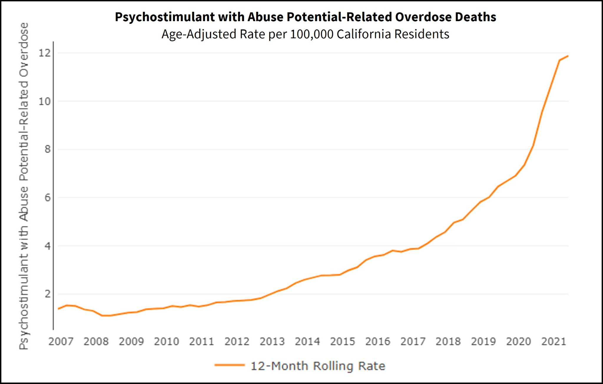
Did you know that amphetamine-type stimulants are the most widely used drugs in the world after cannabis?  Did you know that  between 2011 and 2016, overdoses from methamphetamine TRIPLED and that 1/4 of all overdoses in 2021 in the US were due to meth?

Methamphetamine use disorder can be seen all over the nation, but prevalence varies per region. Rates are highest in the West Coast and South. For example, prevalence of reported meth use in the past year in CA is reported at 1.04% of all adults, almost twice as much as most states in  the Northeast (see map below).
 
In California, non-fatal ED visits and overdose deaths have both risen over the last decade. In fact 32% of those in court-mandated substance use disorder treatment programs were there due to methamphetamine use. While the bulk of media and political attention is currently focused on opiates, one wonders, why aren't we talking more publically about methamphetamine?


What is methamphetamine?
Methamphetamine is an amphetamine derivative, notable for its additional methyl group; it enhances dopamine and norepinephrine in the synaptic cleft. Meth has a very long half life (12 hours cmpared to 90 minutes for cocaine). 

Why is meth bad? So many reasons. . . keep reading to understand a few of the major adverse effects. 

Cardiovascular toxicity

CV toxicity is the #1 cause of death in patients using methamphetamines, and risk of sudden cardiac death is increased by 27% with active meth use. CV toxicity includes a range of end-organ issues, including:
1) Hemorrhagic and ischemic strokes, due to vasoconstrictive effects and cerebral hypoperfusion
2) Very high rates of coronary artery disease (CAD) -- half of patients with regular meth use have CAD, despite lower rates of obesity and diabetes in these patients. This is thought to be directly related to the pro-inflammatory effects of meth. 
3) Angina, which does not respond well to nitroglycerin, is common, due to vasospasm
4) Pulmonary hypertension, especially with IV meth use, due to damage to pulmonary endothelial cells
5) Severe systolic dysfunction with LV dysfunction is another sequalae of meth use
6) Ventricular arrhythmias are notable

Neurotoxicity
Neurotoxicity is the #2 cause of morbidity and mortality in patients using meth. It rapidly crosses the blood brain barrier. It does a doozy on the brain, including disrupting pleasure centers, creating episodic memory issues, damaging executive function (2/3 of people with regular meth use show cognitive impairment, worse with older age and longer duration and frequency of use), disrupting motor function (including fine motor and choreas), and can lead to psychosis similar to schizophrenia (delusions of persecution, auditory hallucinations, and formication in almost half of people using). 

There is also a direct relationship between meth use and Parkinson's disease.

Dental effects
Serious dental effects include caries, tooth loss, tooth fractures -- all due to decreased saliva production (xerostomia), teeth grinding and jaw clenching that occurs with meth use.

Medication Assisted Therapy (MAT)?
Unfortunately, there are no FDA approved treatments for methamphetamine use disorder. A large meta-analysis of 43 RCTs with over 4000 patients found no clear evidence-based effective treatment. 

These included trials with mirtazapine (conflicting results), methylphenidate, bupropion, naltrexone and modafinil (limited evidence of benefit, no support for routine use). In addition, anticonvulsants, antidepressants, antipsychotics all low strength and insufficient evidence. Bummer. 

There was a small study that suggests that methylphenidate may be associated with decreased use over time: no difference at 30 days, but decreased in self reported use days at 10 weeks. 

Also, a small study of combination therapy --  IM naltrexone (380mg q3 weeks) PLUS PO bupropion (450mg daily) small treatment effect of 11% reduction in meth use. 

Hopefully, people will continue to investigate different agents for MAT and treatment of meth use disorder!

In conclusion, Dr. Freschl recommended that we use shared decision-making with patients when talking about trialing non-FDA approved treatment options. She reminded us to screen for CV and neurological sequelae of methamphetamine use. 

Pulmonary Hypertension (Wang - 8/2/23)

 A recording of this presentation can be viewed HERE.

***

Dr. Helena Wang gave an excellent Grand Rounds this week on Pulmonary Hypertension. It is worth watching!

We currently have at least three patients on our adult medicine service that we are evaluating for this condition after getting a TTE to look at their heart failure status. Pulmonary hypertension is such an interesting intersection between the lungs and the heart and a good opportunity to review basic physiology too! Dr. Wang gave an earlier version of this presentation 2 1/2 years ago, and I felt then, as I did this week, that I could watch it over and over. You can! 

But for those who prefer the written word:

The definition of pulmonary hypertension (pHtn) is simple enough: a mean PAP of > 20mmHg at rest.

Dr. Wang likens the pulmonary artery to something like a six lane highway (think of the 101 going north from Novato into Petaluma). When the lanes decrease from six to two in northern Novato, there is suddenly a lot of congestion, and all the cardiac output has to move through that smaller space at higher pressures. Voila! Elevated PAP.

Pulmonary arterial hypertension is rare, but not that rare, 15-50 cases/million, and more common in people with HIV (regardless of CD4 count or duration), connective tissue disease, and portal hypertension. For patients with any of these diagnoses, should be screened annually for pHtn with an echocardiogram (TTE).

Symptoms of pHtn include dyspnea on exertion, fatigue, edema, bloating, and syncope (obviously pretty non-specific). 

On exam, you can see elevated JVP (check the neck veins!), hear a split S2, and see peripheral edema.

Chest imaging reveals often a dilated R pulmonary artery on CXR, and even sometimes a prominent L pulmonary artery. Also, look for the "banana and egg sign" on CT, in which the aortic arch and the pulmonary artery are a little too close in size for comfort. The aortic arch is the banana; the pulmnoary artery is the egg.

Banana and egg sign (photo credit: CHEST Journal)


Okay, what about the TTE??

Oh, reminds the pulmonologist and the family physician, don't forget to pay attention to the right side of the heart in the echocardiogram! Dr. Wang urged us to scroll down, skip the conclusion,  and look at the text (often the third paragraph). Is the RV big? What is the velocity of the regurgitant jet? What is the TAPSE?

A first hint of pulmonary hypertension on a TTE comes on apical 4 chamber view where the R ventricle shows turbulent flow back into the R atrium

RVSP (right ventricular systolic pressure) is an estimate reported on most echo reports and can be very challenging for the Echo tech to calculate. However, a calculated RVSP>25 is considered HIGH right sided pressure and increases your suspicion for pHtn.

The R ventricle is often very plump and dilated under high pressure (so. . .a big chubby RV on echo is another clue that you might have pulmonary hypertension)

Tricuspid Annular Plane Systolic Excursion (TAPSE): RV contracts in a twisting motion with each contraction as RV shortens. Normal excursion is 1.5cm. If RV is volume overloaded and dilated, cannot squeeze as well, not shortening, TAPSE drops <1.5cm on echo

And finally, right heart catheterization is still gold standard for measuring R sided pressures, but really this should be done after you have done a full evaluation (see below) once you are pretty certain you have primary pulmonary hypertension (WHO Group 1). And, due to changes in insurance, it is no longer required always for diagnosis.


WHO Classification of Pulmonary Hypertension
  • WHO Class 1: Pulmonary ARTERIAL Hypertension (pressure goes up, fluid backs up)
    • WHO Class 1 has the worst outcome: 5 year survival 61%
    • only diagnosed once other causes of pulmonary hypertension are identified and treated
    • Seen in patients with HIV (regardless of viral load, duration), connective tissue disease (e.g. scleroderma), and portal hypertension
  • All other WHO classes are actually pulmonary VENOUS hypertension (something downstream to the lungs causing poor forward flow, back up of blood, then high pressure on R side of heart). These include
    • WHO 2: L heart disease (LV systolic or diastolic dysfunction, valvular disease, s/p TAVR, myxomatous disease)
    • WHO 3: bad lung disease or hypoxia (COPD, inadequately treated asthma, ILD, sleep disordered breathing (OSA), OHS, chronic exposure to high altitute, developmental lung disease. Consider high resolution CT (with very thin cuts with and without contrast), home sleep test, pulmonary function tests.
    • WHO 4: chronic thromboembolic pulmonary hypertension (small, little tiny chronic). You cannot see this on typical PE protocol CT scan. You need a VQ scan to evaluate chronic thromboembolic disease.
    • WHO 5: potpourri
Once Pulmonary Hypertension is identified, the next question is DOES the patient have pulmonary hypertension that is primary or secondary

Ddx pulmonary arterial hypertension (WHO Group 1)
  • idiopathic
  • heritable (no current gene therapies)
  • drug and toxin induced (fen fen, methamphetamines including stimulant medications prescribed)
  • connective tissue disease (scleroderma)
  • HIV
  • portal hypertension
  • congenital heart disease
  • Schistosomiasis (#1 cause of pulmonary hypertension outside the US)
A quick interruption to review the three types of CT chest you might consider to evaluate the lungs:
  • CT angiogram (aka CTA) is a very quick scan from feet to head chasing the dye, itskips very low bases and apices of lungs and should never be used to assess for interstitial lung disease. Great for acute PE asessment.
  • High Resolution CT Chest: much thinner cuts (2mm) than a CTA starting at apices and going all the way to the bases. 
    • Very high detail of parenchyma
    • Ground glass opacities can be atelectasis vs. ILD--> flip when prone to see if atelectasis resolves while doing high res scan
  • CT w/wo contrast (e.g. nodule) 
    • can see 4 generations of branches of bronchial tree, but keep in mind that there are 17, so you cannot see filling defects in the small peripheral vessels

Treatment of Pulmonary Arterial Hypertension (PAP)

  1. First identify if the patient has any WHO 2-5 diagnoses that may be influencing their pHtn and treat them maximally, then repeat TTE to check for PAP
  2. If they still display pHtn on TTE, they now meet criteria for pulmonary arterial hypertension (PAP).
  3. Consider pulmonary vasodilator therapy (obviously in conjunction with pulmonologist). These, while still very expensive, are coming down in price. The meds range currently between $40,000 to $100,000 per year.

When to treat Pulmonary Arterial Hypertension?
There is NO specific number for pressure goals. Look at patients functional class, 6 minute walk, and Echo
Treatment goals:
  • Functional class: symptoms at rest (class IV), normal (class I): goal is 2 or less
  • 6 minute walk test: goal is 400mg in 6 minutes ("please walk as far as you can in 6 minutes", not "as fast as you can walk")
  • BNP: can be elevated in R heart strain, goal is normal
  • Echo: goal is RV not fat and chubby, decompressed and slender, TAPSE moving nicely
Pulmonary Hypertension Therapy

  • Endothelin pathway: block (bosentan (black box liver toxicity, no longer used), ambrisentan, macicentan: side effects: edema, check LFTs, watch for rare malignant facial edema
  • Nitric oxide pathway: enhance (sildenafil TID, tadalafil QD, no lab monitoring, can get blue green colorblindness, rare complication anterior ischemic optic neuropathy (sudden blindness one eye, reversible when stop), riociguat (TID, more systemic hypotension)
  • Prostacyclin pathway: enhance (potent, short half life and unstable)
    • epoprostenol: IV infusion, tubing connected to a pump, best mortality data, very labor intensive
    • treprostinil: IV vs. sq (like insulin pump) and inhaled QID (takes a lot of time, breathing treatment like nebulizer, 20-30 minutes per treatment
    • iloprost: inhaled 6-9 times/day
    • selexipag: oral BID, no labs but unrealistic uptitration protocol
Guidelines for treatment are based on functional class. The ultimate goal is minimal symptoms with regular activity
If functional class IV-->  needs prostacyclins right away
If functional class III-->  can start with orals (e.g. tadalafil and macicentan)

Any questions? Email or staff message Dr. Helena Wang at: helena.wang@sutterhealth.org


Diagnosis and Management of Osteoporosis (Hamann 7/23/2026)

 A recording of this presentation is available HERE .  *** Thanks so much to Dr. Kendal Hamann, SMGR Endocrinologist, for an outstanding Gra...